Coronaviruses 👑 (Coronaviridae) 1968
Coronaviruses (Coronaviridae) are a family of viruses that combines medium-sized RNA-containing pleomorphic viruses, on the surface of which there are characteristic fringed villi. Initially, the viruses belonging to this family were classified as myxoviruses, but the results of subsequent studies gave grounds (1968) to separate them into an independent taxonomic group. The generic division of the Coronavirus family has not been determined.
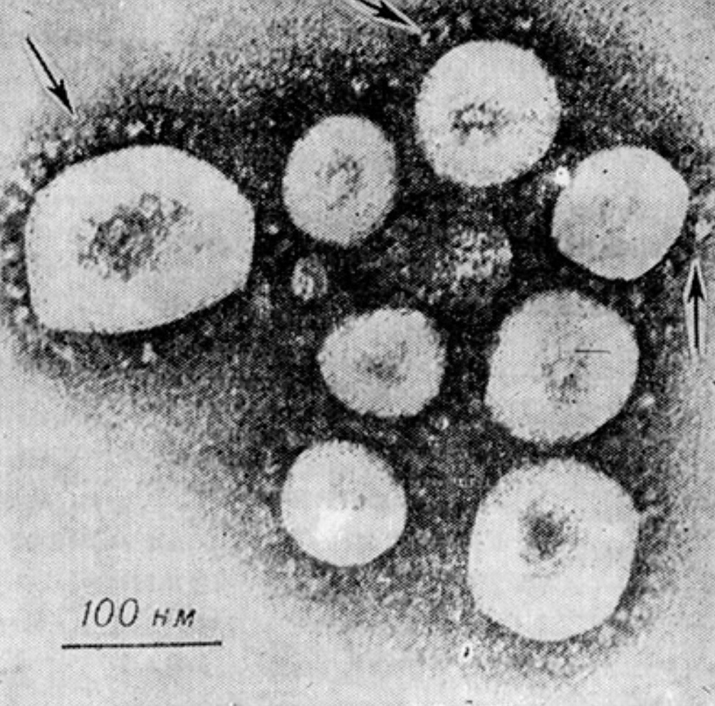
Electronogram of cytoplasm of W1-38 cells infected with human coronavirus, strain 229 E: synthesized sickle-shaped structures (1) and budding virions (2) of coronaviruses are visible in the region of large vacuoles (K. McIntosh, 1974).
Coronaviruses multiply in the cytoplasm of infected cells. In this case, daughter virions appear after 4-6 hours. after infection, and the maximum manifestation of cell-associated infectivity is noted after 12-36 hours. In the electron microscopic examination of infected cells, a rather characteristic morphology of K. is revealed: near the cytoplasmic vacuoles, the appearance of sickle-shaped structures, which are synthesized virions, is observed, and budding virions appear in the vacuole itself (Fig. 2). Budding through cytoplasmic vacuoles is a characteristic feature of K., which distinguishes them from myxoviruses that bud through the plasma membrane.
The type species of K. is considered to be a virus of infectious bronchitis of birds. The K. family also includes human c. - respiratory viruses, hepatitis mouse virus, transmissible gastroenteritis virus in pigs, rat sialodacryoadenitis virus, turkey blue scallop virus, neonatal calf diarrhea virus, and hemagglutinating swine encephalomyelitis virus. According to antigenic properties, these viruses are heterogeneous. In the immunodiffusion test, they were able to identify one to four lines of precipitation. However, there are some antigenic links between certain human (OS 43 serotype) and mouse strains.
All K. inherent in the ability to fix complement in the presence of hyperimmune serums or serums obtained from patients.
Only two types of Coronaviruses (Human Coronavirus, STRAINS OS 38 and OS 43 and Hemagglutinating Swine Encephalomyelitis Virus) cause hemagglutination; at the same time, human erythrocytes agglutinate human and monkey erythrocytes only at t° 4°, whereas the erythrocytes of chickens, rats and mice agglutinate both at t° 4°, and at room temperature and at t° 37°. The ability to cause hemagglutination was impaired after treatment with ether and trypsin. Moreover, it is shown that as a result of exposure to the named K. bromelain, which destroys fringed villi, there was not only a violation of hemagglutination, but also a violation of complement fixing and inf. Activity K.
Coronaviruses can multiply only in a limited number of cell cultures, and when using cells grown in the form of a monolayer, it is possible to obtain a very low yield of the virus. K. of various origins reproduce, as a rule, only in the cells corresponding to their origin. Thus, the primary isolated bird plants reproduce only in the cells of birds, mouse cats reproduce only in the tissues of mice; the same can be said of rat and pig strains. In chicken embryos, only the C. of infectious bronchitis of birds reproduces. Human cells reproduce, exhibiting strain differences, in human diploid cells, in L-132 cells (a cell line derived from a light human embryo), as well as in HeLa cells, cells of the primary culture of the kidney of a human embryo, and other human cells. Reproduction of some viruses can be detected only in organ cultures (e.g., reproduction of the human virus in the explantates of the epithelium of the trachea of the human embryo). Cytological methods do not detect any inclusions in the affected cells. It should be emphasized that the cytopathic effect of K is not manifested in all cases. Some viruses (e.g., human C. strain B814) can only be detected by an electron microscope or by interference with other viruses (for the human C. named as an example, such an interfering virus is an ECHO virus of type 11). When cells were coated with methylcellulose, plaque formation was observed in L-132 and Wl-38 cell cultures.
In the experiment, it was possible to achieve the adaptation of Coronaviruses to cells in which they were not primarily reproduced. So, the synthesis of the virus inf. bronchitis of birds was observed in the primary culture of monkey kidney cells, and human kidney cell culture was observed in monkey kidney cell culture and the body of sucking mice.
Human coronaviruses
Human coronaviruses are, as a rule, causative agents of acute diseases of the upper respiratory tract, but there are cases of bronchitis and pneumonia of coronavirus etiology (in children). Accurately establishing the role of this virus in comparison with epidemiological data is extremely difficult due to the wide variability of strains and the complexity of the diagnostic method.
K. is usually isolated from nasal secretions, scrapings and washing waters of the nasopharynx, using appropriate cell or organ cultures. The effectiveness of the isolation of K. is relatively small. Diagnosis is based on virological (electron microscopy of infected cultures, as well as cytopathic action detected in the corresponding monolayer cell culture) and serological (increase in the serum of the patient's number of antibodies to the complement-binding antigen of human C. strain, strain 229 E, and antibodies to hemagglutinin strain OC 43) methods.
Virological studies 🔬
VIROLOGICAL STUDIES are studies conducted to diagnose viral infections, study the relevant pathogens, their spread in nature, as well as in the production of viral drugs. In virological laboratories (see) honey. profiles are studied as human viruses, and in some cases animal viruses (e.g., conduct the diagnosis of rabies in dogs, examination of animals used for the production of viral drugs). The methods of studying both are similar.
One of the main stages of virus is the isolation of viruses. When isolating viruses from people, blood, various secretions and excreta, pieces of organs are used. Most often, the blood is examined for arbovirus diseases. Whole defibrinated or Hemolysized blood, its individual elements or clots (in the later stages of the disease) are used. Viruses of rabies, epid, mumps, herpes simplex can be found in saliva. Nasopharyngeal flues serve to isolate pathogens of influenza, measles, psittacosis, rhinoviruses, respiratory syncytial virus, adenoviruses. Adenoviruses are also found in washes from the conjunctiva. Flush is taken by rinsing the nose and pharynx (separately) and washing the conjunctiva with an isotonic solution of sodium chloride. You can wipe the nasal passages and the back wall of the pharynx with tampons moistened with broth. Non-sterile material is treated with antibiotics (1000 IU of penicillin and streptomycin per 1 ml) for 30 minutes. Samples are diluted 1:10 with a phosphate buffer, centrifuged twice for 20 minutes. at 8000 about I min. Antibiotics are added, as indicated above. Less often for V. I. take the contents of pustules (with smallpox, chickenpox, herpes) and organ punctates (with venereal lymphogranuloma). Sectional material should be taken as soon as possible after the death of the body. It is stored until the moment of study at t° -20 ° and below. To carry out the V. I. the tissue is crushed (rubbed) and 10-20% suspension is prepared on an isotonic solution of sodium chloride or a nutrient medium for cell cultures. It is centrifuged for 20 minutes at 1500 rpm; supernatant fluid is used for further research.
In order to isolate viruses, laboratory animals, bird embryos, cell and tissue cultures are infected. Animals are suitable if the virus causes them clear clinical symptoms of the disease or pathoanatomical changes (e.g., paralysis, pneumonia, etc.). From the tropism of the virus depends on the effectiveness of a particular way of introducing the material. Widely used infection under the skin, intraperitoneally and intravenously. Neurotropic viruses are detected when infecting animals in the hemispheres of the brain (arboviruses, rabies virus, etc.), visual tubercle (polio virus in experiments on monkeys), spinal cord. Smallpox and herpes viruses can be detected by applying material to rabbits on the scarified cornea. Some viruses are easily detected by inoculation into the ingerierio chamber of the eye (e.g., dog hepatitis virus in puppy experiments). To study the causative agents of respiratory infections, intranasal infection of animals is usually used (instillation of material into the nose of a narcotized animal or its introduction in the form of an aerosol in a special chamber). In the digestive tract, the material is injected with food or through the mouth with a blunt needle. In the study of some oncogenic viruses, the method of infection of golden hamriches into the mucous membrane of the cheek sacs is used.
To many viruses, newborn animals and suckers are more susceptible than sexually mature individuals. Sucking mice are widely used to isolate arboviruses and Coxsackie viruses (after infection in the brain). Some adenoviruses are able to induce tumors in subcutaneous infection of newborn golden hammags. The study of a number of bird viruses is carried out on chickens of the first days of life.
Rice. 1. Pockpockets of the vaccine virus on the chorion-allantois shell of a chicken embryo.
Rice. 1. Pockpockets of the vaccine virus on the chorion-allantois shell of a chicken embryo.
The use of chicken embryos has a number of advantages. Their undifferentiated tissues have a wide range of sensitivity to many viruses. The presence of infection is judged by the death of embryos, the appearance of changes (pockmarks) on the chorion-allantois membrane (Fig. 1), the accumulation of hemagglutinins and complement-binding viral antigen in embryonic fluids. Infect embryos on the chorionallantois membrane (at the age of 11 - 12 days with viruses of the smallpox group), in the allantois and amniotic cavities (10-11-day myxoviruses), the yolk sac (at the age of 5-6 days by the causative agents of psittacosaoornitosis, etc.). Inoculation of the material to embryos in the brain and intravenously (in the vessels of the membranes) is rarely performed. With any method of infection, embryos can be injured, so they died in the first 24-48 hours. excluded from accounting.
To study the effect on chemical viruses. Deembryonic eggs are very convenient, in which the embryo is removed, but the chorionalanthis membrane is preserved. Inside place the virus and the substance under study in 20 ml of isotonic sodium chloride solution. The hole in the shell is closed with a cap with a tube, through which samples can be taken for analysis.
When evaluating experiments on animals and embryos of birds, it should be borne in mind the possibility of provoking latent infections in them or isolating a virus in a latent state.
Exceptionally widely for the isolation and accumulation of viruses used cultures of cells and tissues (see). These methods can cultivate most of the known viruses (see Cultivation of viruses). Some of them intensively accumulate already during the initial infection of crops, for the adaptation of others requires several passages. The reproduction of most viruses in cell cultures is accompanied by the development of a cytopathic effect. By its nature, to a certain extent, it is possible to judge the belonging of viruses to one kind or another: picornaviruses cause rounding and wrinkling of cells, adenoviruses - the formation of clusters by rounded cells in the form of grape clusters, myxoviruses and herpetic viruses - the formation of multinuclear syncytiums. A number of viruses can not be cultivated outside the body.
The reproduction of some viruses (smallpox group, myxo- and arboviruses) can be detected by a hemadsorption reaction, since the affected cells acquire the ability to adsorb red blood cells. The corresponding erythrocytes (humans, monkeys, guinea pigs, chickens) in a concentration of 0.4-0.5% are placed on a monolayer at t° 4 ° or at room temperature for 20-30 minutes.
The reproduction of the virus is sometimes judged by studying the culture fluid in animals (tick-borne encephalitis) or in the RSC. The presence of a virus that does not have cytopathic activity is sometimes determined by its ability to interfere with the cytopathogenic virus. Thus, in the cell cultures of chicken embryos infected with bird leukemia viruses, the reproduction of the Rouse sarcoma virus is suppressed. To detect non-cytopathogenic strains of bovine diarrhea viruses and swine cholera, the END (exaltation of Newcastle disease virus) method is proposed - superinfication of cultures with the Newcastle disease virus. With the joint action of both viruses, cell destruction occurs.
When cytopathic changes or other signs of virus reproduction appear, the culture fluid is used to identify the virus or passage. A number of viruses remain associated with cells even with culture degeneration (adenoviruses, smallpox viruses), as a result of which they freeze and thaw cultures before collecting fluid. Some herpetic viruses, such as Marek's disease virus in chickens, need to be reseeded along with intact cells.
To study human respiratory corona viruses and some others, the method of tissue cultures is used, i.e. infection of tissue fragments cultivated in vitro. Most often use the tissue of the trachea of a rabbit. The multiplying virus affects the cells of the endothelium of the mucous membrane, which is determined by stopping the movement of the cilia.
The possibility of the presence of foreign viruses in tissue and cell cultures should be taken into account. They can be introduced with cells, if the latter are taken from an infected organism, get from trypsin or serum used to culture cells.
In addition to sowing biopsy or sectional material on already grown crops, direct cultivation of cells of the organ under study after its trypsinization is used, which is often more effective with regard to the isolation of the virus (for example, the detection of adenoviruses in the tonsils). The technique of mixed cultures is also used, when the cells of the organ under study are grown together with any cells sensitive to this virus (for example, seeding of brain cells of patients with subacute sclerosing panencephalitis together with monkey kidney cells or Hela cells to isolate the measles virus). The method of mixed cultures is often the only way to isolate the virus from tumors induced by it in animals that do not produce an active virus, but contain a viral genome.
Rice. 2. Plaques of polio virus on monkey kidney cell culture.
Single-layer cell cultures make it possible to obtain colonies of the virus - plaques (Fig. 2). As a rule, plaques form viruses that have cytopathic activity. At the same time, this method makes it possible to detect some non-cytopathogenic viruses (for example, a number of strains of bovine diarrhea virus). To obtain plaques, the virus is introduced on a cell monolayer in cups or flat vials. The multiplicity of infection, i.e. the number of viral particles per cell, should be small so that the plaques formed do not merge. After 30-60 min. adsorption, the nutrient medium is layered with 1.35 - 1.5% agar and neutral red in the final dilution of 1: 40 000. Cultures in petri dishes are incubated in the atmosphere with 5-10% carbon dioxide, and hermetically sealed vials - in a conventional thermostat. After a few days, uncolored foss of degenerated cells begin to stand out among the lifetime-colored cells.
You can place agar on the cells without a neutral red, and after a few days apply a second layer of agar with dye; plaques become visible after a few hours. Agar sometimes contains polysaccharide sulfates, which are inhibitors of virus growth; to neutralize them, protamine sulfate (60 mg per 100 ml) is added to the medium. To obtain plaques of a number of viruses, methylcellulose and other substances can be used as a coating. Some viruses (smallpox, measles) form plaques without agar coating. The plaque method allows for clonal analysis of viral strains. To isolate genetically homogeneous clones, one plaque is extracted, which is used for the next infection. Usually cloning is carried out over three passages.
The plaque method is also suitable for determining in an infected culture the number of cells that produce the virus (i.e., the number of infection centers). To do this, the cells are suspended, placed on a single-layer culture of virus-sensitive indicator cells and poured with agar. Plaques form around the infected cells.
To diagnose viral infections and study the antigenic structure of viruses, a precipitation reaction in the gel is used. Most often, agar is used for this purpose. Antigens and specific antibodies placed in an agar gel at a certain distance diffuse and form a precipitate in the form of white stripes when they meet. 0.8-1% agar in an isotonic solution of sodium chloride or phosphate buffer is placed in capillaries or applied in a layer on slides. Antigens preferably have purified and concentrated. The reaction ingredients are applied to the agar in the opposite ends of the capillary or in the holes made in the agar layer on the glasses at a distance of 5-6 mm. Incubation lasts 4-20 hours.
A significant number of V. I. perform with the help of light and electron microscopy. The largest viruses (e.g. smallpox) after appropriate treatment (silvering, Victoriablau staining, etc.) can be detected by conventional light microscopy. This method is used in the diagnosis of smallpox by examining the material from pustules. Characteristic of some infections is the formation in the cells of bodies - inclusions. So, in the nuclei there are inclusions in herpetic and adenovirus infection, in the cytoplasm - with smallpox (Guarnieri bodies) and rabies (Babes-Negri bodies). Detection of inclusions is important for the diagnosis of rabies, smallpox, cytomegaly, subacute sclerosing panencephalitis, etc.
Microscopy in a dark field (see Dark Field Microscopy) and phase-contrast microscopy (see) are used to study the dynamics of changes in the cells affected by the virus. More widely used luminescent microscopy (see).
Examine smears, prints and single-layer cell cultures grown on glasses. Drugs (native or fixed) are most often stained with acridin orange. The method allows you to identify large viruses and clusters of viral components. Formations containing DNA glow bright green, and THOSE containing RNA glow brick-red. Even more often, when V. I. produce treatment of infected cells with fluorescent antibodies, which allows you to identify accumulations of viral antigen. With the direct method, immune gamma globulin labeled with a fluorescent dye is used, for example, fluorescein-isothiocyanate. With the indirect method, the drug is treated with the usual immune serum of an animal, and then labeled antibodies against the gamma globulin of this animal. Drugs are viewed in ultraviolet light, viral antigen is detected by a light green glow .
The method of smears from the nasopharynx allows for early diagnosis of respiratory viral infections - influenza, parainfluenna, rhino- and adenovirus, respiratory syncytial.
Electron microscopy (see) allows you to study the size and structure of viral particles, as well as the subtle changes caused by them in cells. Examine viral suspension or ultra-thin sections of infected cells. Some viruses (e.g., a number of human and animal oncornaviruses) can be detected in tissues only by this method.
V. I., the purpose of which is to determine the number (titer) of the virus or viral antibodies, are very diverse.
Under an electron microscope, you can count the number of viral particles in the suspension, if it is sufficiently cleaned. The virus is mixed with polystyrene latex, the number of particles to-rogo is known. The mixture is sprayed on a grid, the number of particles in individual drops is counted. According to the ratio of the number of latex particles and viruses, the number of virions in a given volume is detected. This method does not give an idea of the infectious activity of the virus, since viral suspension usually contains a significant number of non-infectious particles.
The most accurate to determine the number of infectious units in the material allows the method of plaques. Single-layer cell cultures infect with a small dose of the virus. The titer is expressed by the number of plaque-forming units (BOE) in 1 ml. Similarly, it is possible to determine the titer of some oncogenic viruses by the foci of transformation, as well as those pathogens that form pockpockets on the chorionallantois shell of chicken embryos (for example, the smallpox virus).
Less accurate is the determination of the titer of the virus by the method of final dilutions. Successively increasing dilutions (usually tenfold) are administered to sensitive animals, into chicken embryos or cell cultures. Taking into account the result of each administration as positive or negative, determine the lowest dose of the virus that can cause a certain effect (disease or death of the animal, the appearance of cytopathic changes in culture, etc.). In practice, this dose is difficult to determine, so they calculate the dose that gives a 50% effect (ED50). It is determined depending on the properties of the virus and the titration method by the number of dead animals (LD50), the number of cases (ID50), the number of embryos that have viral hemagglutinins or complement-binding antigens, the number of cell cultures where cytopathic changes or hemadsorbing activity were detected. The titer is expressed by the number ED50 in a certain volume of material.
Of the existing methods of calculating ED50, the most common is the Reed-Munch method, based on the principle of cumulation. It is based on the assumption that each test object (mouse, embryo) that responded positively to the introduction of this dilution would respond positively to the introduction of a more concentrated virus. And, conversely, if this dilution caused a negative reaction, then with the introduction of more dilution, there will also be a negative reaction. The intervals between dilutions with this method should be the same, each breeding infects at least 4 animals (cultures). Next, interpolation is performed between doses that caused the effect closest to 50%.
Where B is the lowest dose that caused the effect of more than 50%, b is the effect of dose B in percentage, and is the effect of the highest dose that caused the effect of less than 50%, in percentage, d is the interval between the logarithms of the two adjacent doses.
Viruses with pronounced cytopathic activity (e.g., polio virus) can be tited by color testing. It is based on the fact that in the process of cell growth, the pH of the environment decreases, and in infected cultures where cells degenerate, changes in the pH of the environment do not occur. To detect these changes, an indicator is added to the environment - phenol red, which has a red color at a pH above 7, orange at a pH of 7 and yellow at a pH below 7. In a nutrient medium having a pH of 7.3-7.4, phenol red is introduced in a final concentration of 1 : 40,000. Determine the minimum dose of cells that changes the color of the medium from red to yellow (usually about 25,000 in 0.25 ml). Next, prepare 10-fold dilutions of the virus, each of which is poured into 4-6 test tubes, where cell suspension is added. Test tubes are closed with rubber stoppers or pour 0.6-0.8 ml of vaseline oil. The results are taken into account after aging for 5-7 days at t° 37 °. For the titer of the virus take its dilution, preventing the shift of pH to the acidic side in 50% of test tubes.
The ability of immune sera to neutralize the infectious activity of viruses is determined by a neutralization reaction. Methods of titration of serums are predetermined by methods of titration of the corresponding viruses. All of them are based on the preparation of mixtures of the virus with serum, which are then tested for the presence of a non-neutralized virus. The reaction is put in two versions. According to one of them, the immune and normal serum in one dilution (e.g., 1: 10) is mixed with increasing 10-fold dilutions of the virus in equal volume and the titer of the virus in the presence of both serums is determined. The partial division of the ED50 virus in the presence of normal serum into its ED50 in the presence of the immune system will be the neutralization index of this immune serum dilution. It is impossible to interpret this result for other serum dilutions (for example, the activity of undiluted serum will be higher than calculated). In another variant of the reaction, a constant dose of the virus (usually about 100 ED50) is combined with a number of 2-fold dilutions of the test serum. The titer of the serum will be diluted, with which the virus caused a 50% effect.
Depending on the method of titration of the virus, the neutralization reaction is carried out on laboratory animals, chicken embryos (according to their death or accumulation of hemagglutinins), on cell cultures (by the appearance of cytopathic changes, color sample, etc.). If the virus forms pockmarks on the chorional anthoise shell of chicken embryos, determine the greatest dilution of serum, reducing the number of pockmarks by 50 percent or more. Similarly, serums are titrated by reducing the number of viral plaques on single-layer cell cultures.
Titration of viruses according to RGA and antibodies according to RTGA is based on the ability of a number of viruses (myxoviruses, some representatives of the genus of poxviruses, adenoviruses, picornaviruses and togaviruses) to glue red blood cells. The virus is titrated in double dilutions with an equal volume of 0.5-1% of the suspension of red blood cells. For a titer (1 AE - agglutinating unit) take its greatest dilution, which caused hemagglutination (see). The hemagglutinating titer of the virus is not an indicator of its infectious activity, since hemagglutinations) can cause non-infectious "incomplete" particles, an inactivated virus and a hemagglutinating antigen that is separated from some viruses. Serums according to RTGA are titrated in double dilutions with 2-4 AE of the virus; the titer is considered the greatest dilution, completely inhibiting hemagglutination).
Some viruses are adsorbed on red blood cells, but do not cause their agglutination. To identify them, you can use the indirect hemagglutination reaction. It is based on the agglutination of red blood cells "loaded" with the virus with serum specific to this virus. This method is used mainly for titration of serums.
The complement binding reaction (see) is suitable for titration of both viruses (more precisely complement-binding viral antigens) and antibodies.
In principle, RSC with viruses does not differ from that with bacterial and other antigens. Differences exist only in the method of preparation of antigens. As viral antigens can serve as blood, nasopharyngeal flues, urine, feces of patients, suspensions of infected organs, individual parts of infected chicken embryos and the virus grown in cell cultures. The most suitable cultural antigens. Suspensions of infected organs before use are repeatedly frozen and thawed, combined with an equal volume of ether or chloroform, shaken for 1-2 hours, after which they are kept for 18-20 hours. at t° 4°, re-freeze, thaw and lighten by centrifugation. To obtain serums, animals should not be immunized with a virus cultured in the same substrate as the reaction antigen. Depending on the purpose of titration, double dilutions of the antigen with a certain dose of serum or, conversely, double dilutions of serum with one dose of antigen are combined. Contact of antigen, serum and complement is carried out within 1 hour at t° 37 ° or 18 hours. at t° 4—6°. After adding the hemosystem, the material is incubated for 30 minutes at t° 37°. Evaluation of the results is carried out according to the general rules.
There are methods of titration of viruses and antibodies based on the indication of the virus in infected culture cells with fluorescent antibodies, as well as a number of others that are used quite rarely.
The toxic effect inherent in certain viruses is detected by administering large doses to animals in an unusual way, with which the virus does not undergo a full cycle of reproduction. So, for example, the flu virus is injected into the brain or intravenously. Its toxic effect is judged by the death of animals during the first day.
The antigenic activity of the virus (or vaccine) is established by immunizing people or animals, followed by determining the antibody titer in their serum. The immunogenic activity of the vaccine, i.e. the ability to cause resistance to infection, is determined by immunization of animals sensitive to this virus. The immunized animals and control non-immunized are then infected with a number of virus dilutions, identifying its ED50 for both groups. The difference in the obtained indicators characterizes the immunogenicity of the tested drug. If the virus is not pathogenic to animals, the immunogenicity of the corresponding vaccine can be determined only in the epidyrol. Experience.
To study a number of properties of viruses (size, structure, chemical composition, etc.), it is necessary to have a purified material with a high titer. Most often, a virus grown in cell cultures or in the form of allantois fluid of infected chicken embryos is used. Purification usually begins with differential centrifugation: first, at 15-20 thousand g (g - acceleration of gravity), the material is released from cellular detritus, and at 50-100 thousand g - the virus itself is besieged. To get rid of large particles, filtration through asbestos gaskets (Seitz type), glass and cellulose membrane filters (such as millipore filters) are also used. Fragments that are smaller than viruses can be removed by filtering the material through sefadex gels, which delay particles of a certain size depending on the pore size. To isolate and concentrate the virus from large volumes, salinization with ammonium and sodium sulfates, precipitation with alcohol, aluminum hydroxide or polyethylene glycol are used. To release ballast proteins, their digestion by proteolytic enzymes is also used. Myxoviruses can be purified and concentrated by adsorption on red blood cells at a low temperature with eluction at a higher temperature.
Further purification and concentration of the virus is carried out by one or another method of fractionation. These methods are also used to separate virus mutants and individual viral components. When mixing the virus with a phase system formed by aqueous solutions of two polymers, it passes into one of the phases depending on its size, ionic composition of the medium, pH, etc. To purify large viruses, the dextran sulfate system - methylcellulose is usually used, small - dextran sulfate with polyethylene glycol. Polymers left over from phase separation are often removed during further purification of the virus. You can also remove dextrasulfate by adding potassium chloride, methylcellulose with ammonium sulfate, polyethylene glycol chloroform and other methods.
Purification of viruses using chromatography on the column is based on their ability to adsorb on a number of substances, and when changing the pH or salt concentration, elutate from them (see Chromatography). For adsorption, calcium phosphate gels, cellulose-based ion exchangers (most often anion exchangers, for example, diethylatminoethylcellulose), ion exchange resins are used. The ellution of the virus from calcium phosphate is carried out by phosphate buffer solutions of increasing concentration, and from cellulose-based ion exchangers - with sodium chloride solutions.
Highly purified suspensions are obtained by centrifugation in a column of liquid with a density distributed in a continuous gradient. Viral particles collect at a level where the density of the medium is equal to their floating density. Zonal centrifugation in the sucrose gradient allows the particles with different masses to be separated. The material is applied to the gradient created by layering the appropriate solutions of sucrose. At the end of centrifugation, fractions are collected, piercing the bottom of the test tube. In equilibrium or isopic centrifugation, viral particles are suspended in a solution of cesium chloride, cesium sulfate or rubidium chloride. After prolonged centrifugation, a stable continuous gradient of the density of the solution is created. At the place of localization of viral particles, their density can be determined. It should be noted that the obtained values of the density and mass of viral particles to a certain extent depend on the medium used in centrifugation.
Viruses labeled with various radioactive isotopes are widely used in the virus. The most commonly used carbon is 14C, tritium 3H, phosphorus 32P, sulfur 35S, iodine 131I. It is easiest to mark the virus when it is cultured in cell cultures.
Chem. the composition of viruses is investigated by generally accepted chemicals. Methods. Nucleic k-tu is usually obtained by phenolic extraction, anionic detergents are less often used - sodium dodecyl or lauryl sulfate.
To identify viruses (see) first of all, it is necessary to establish their generic affiliation.
To do this, it is necessary to determine the size and structure of viral particles, the type of nucleic acid included in their composition, the presence of a lipoid shell. The type of nucleic k-you is most often determined by indirect methods, for example, using the ability of bromodeoxyuridine to suppress the reproduction of DNA-containing viruses. The presence of a lipoid shell in the virus is established by its sensitivity to the action of ether and chloroform (viruses with a shell are inactivated). Further identification is carried out with a set of immune serums to known viruses, using various reactions - neutralization, RSC, RTGA, etc. Less often immunize animals with a known virus with their further infection with an unknown or vice versa.
Bibliography:
Laboratory diagnosis of viral and rickettsial diseases, edited by E. Lennet and N. Schmidt, trans. from English, M., 1974, bibliography;
Luria G. E. and Darnell J. E. General Virology, trans. from English, Moscow, 1970, bibliography; Methods of Virology and Molecular Biology, trans. from English, Moscow, 1972; Pshenichnov V. A., Semenov B. F. and Zezerov E. G. Standardization of methods of virological research, M., 1974, bibliography; Manual of Laboratory Diagnostics of Viral and Rickettsial Diseases, edited by P. F. Zdrodovsky and M. I. Sokolov, M., 1965; Sokolov M. I., Sinitsky A. A. and Remezov P. I. Virological and serological studies in viral infections, L., 1972; Virologische Praxis, hrsg, v. G. Starke, Jena, 1968, Bibliogr.
E. P. Pille.
Category: Volume 4
Source: Great Medical Encyclopedia (BME), edited by Petrovsky B.V., 3rd edition
In most cases, coronavirus causes people to be sick between January and March. The spread of the disease is mainly intra-family.
Vaccine prevention and chemotherapy for coronavirus diseases have not been developed.
Bibliography: Zakstelskaya L. Y. and Shcheboldov A. V. Coronaviruses of man and animals, M., 1977, bibliography; Mc Intosh K. Coronaviruses, Curr. Top. Microbiol. Immunol., v. 63, p. 86, 1974; Tyrrell D. A. a. o. Coronaviruses, Nature (Lond.), v. 220, p. 650, 1968; Tyrrell D. A. a. o. Coronaviridae, Intervirology, y. 5, p. 76, 1975; Viral infections of humans, ed. by A. S. Evans, N. Y., 1976.

https://telegra.ph/Terrorism-and-Communism-class-mathematics-and-the-voice-of-the-proletariat-07-30


Leon Trotsky's great-granddaughter Nora Dolores Volkova-Bronstein, director of the U.S. National Institutes of Health.
These are Soviet Bolshevik knocks 100% ⬇️

Additional materials special operation corona 👑 here 🔜🎙 👇
https://telegra.ph/CONSPIRACY-OF-SILENCE-CROWN-08-30
https://telegra.ph/STATISTICS-ON-THE-FATE-OF-VACCINATED-AND-UNVACCINATED-CHILDREN-08-30
https://telegra.ph/STATE-INTERNATIONAL-BIOLOGICAL-TERRORIST-ACT-CORONA--19-08-31
https://telegra.ph/FALSE-VACCINES--FROM-PFIZER-POISON--FROM-THE-WORLD-CROWN-09-01
https://telegra.ph/INTERNATIONAL-BIOLOGICAL-AGRICULTURAL-TERRORISTS--21-09-01
https://telegra.ph/PERSONALIZATION-OF-THE-COMPOSITION-OF-THE-STATE-CRIMINAL-GROUP-CORONA--21-09-02

ŴξLL, ͲΗΑͲ'Ѕ 𑀡Ͳ, ϺΑLϒΑͲ

@brigadegeneralino_bot