Peter McCullough interprets a cell’s own structures as "viruses"
NEXT LEVEL - Wissen neu gedacht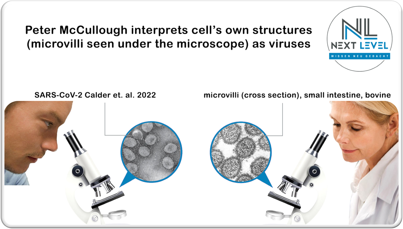
A shock, not unlike an earthquake, swept over the world on Nov. 17, 2022 – especially the international virologist scene. An unparalleled frenzy of joy broke out, for at last! – finally the irrefutable proof of a pathogenic and existent virus has been provided! In the front line here is the US cardiologist Peter McCullough, who with his cited publication "Electron cryotomography of SARS-CoV-2 virions reveals cylinder-shaped particles with a double layer RNP assembly" attempted to refute those who reach an ever-growing audience by stating that there is still not a single scientific proof of the existence of pathogenic viruses.
Peter McCullough's article begins with these words:
Based on this claim, the usual suspects jumped quite cheerfully onto this bandwagon, claiming that all our criticisms would now be cleared from the world once and for all – including educational channels, such as:
● ScienceFiles
● Dr. Bodo Schiffmann
● #WirMachenAuf & Zentrale fuer KindesRechtsVertretung – INFO KANAL (Central Office for Children's Legal Representation - INFO CHANNEL)
● and many more.
Not only does it seem surreal when, after almost 3 years, someone finally managed to present the proof (along the lines of "Better late than never"), on the basis of which we had to endure the measures imposed on us by the "pandemic" – but we have been waiting for this proof of the existence of viruses since 1954!
Before we gradually analyze this publication and dismantle it at the same time, we would like to point out our suspicion that the above-mentioned group of people, who regard such a study as evidence of a pathogenic virus and thus celebrated their "victory" prematurely, either did not read this work – or did not understand it. Or both.
Peter McCullough attributes a specific and distinct disease to Covid-19
Peter McCullough stated:
Before we go into more detail about the scientific publication, the first misconception can already be highlighted here.
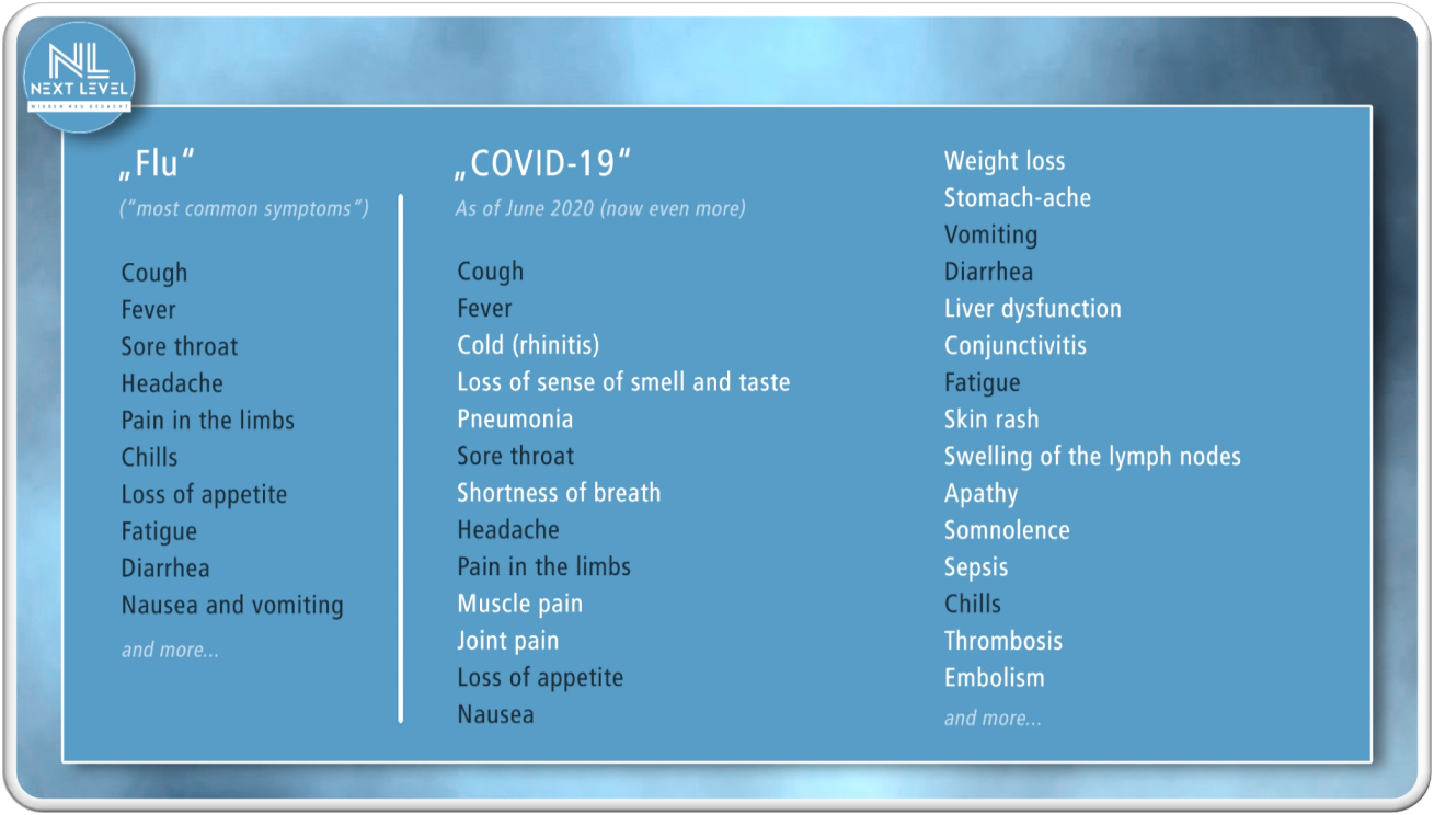
Covid-19 is defined as a "broad, non-specific symptom spectrum". In other words, a bottomless pit. Inevitably, an increasingly variable – precisely non-specific – "spectrum of symptoms" builds up, that is to say a Super-Syndrome Covid (SSC). This means, on the one hand, that the contradictions are becoming more and more obvious, and on the other hand, that they can no longer be concealed or argued away.
If one compares the symptoms attributed to Covid-19 with those of the flu, it is obvious that Peter McCullough's statement is incorrect and that by no means anything specific or new has emerged – the spectrum of symptoms presents itself in an almost unmanageable variety.
Peter McCullough’s proof of the existence of pathogenic viruses
The publication cited by Peter McCullough, in which one can supposedly find the definitive proof of the existence of SARS-CoV-2 and other pathogenic viruses, is in fact mainly only about close-up images and 3D models of different variants of "SARS-CoV-2".
Peter McCullough hopes that the release of these close-up images of SARS-CoV-2 will allow us to move on to the relevant issues. With this, he may actually be helping the vaccine-obsessed and other beneficiaries of the COVID-19 panic to cause further and even greater damage.
By suppressing the fact that the depicted structures are by no means viral particles, he further reinforces the viral narrative and paves the way for the justification of further vaccines, testing procedures and measures.
The alternative news channel ScienceFiles added in its article [3]:
"However, those who do not want to learn, those who want to continue to defend their ongoing belief, as is so common among religious people, against the anomalies and contradictions in reality, those will likely find similar excuses as Pope Urban and his Jesuit stooges who refused to accept the existence of craters on the moon and the moons of Jupiter when Galileo Galilei made improvements to a Dutch telescope that allowed him to see them clearly.
However, anyone who wants to continue denying the existence of viruses must now, according to the work of Calder et al. (2022), explain:
- What it is which can be seen?
- Why is it identifiable as different variants, which are derived by sequencing the genome of what is there, and which are called Alpha, Beta, Delta and Wuhan variants of SARS-CoV-2?
- Why is it present in people who tested positive for SARS-CoV-2 but not in people who tested negative for SARS-CoV-2?
- Why is it found in people who are seriously ill with COVID-19, but not in people who have lung disease but have not tested positive for SARS-CoV-2?
- Why, then, if what is pictured above is present, can a specific clinical picture occur that does not occur if the "somethings" shown are not present?
And going forward, whoever has a problem speaking of viruses can say Grumph to what is demonstrably present, and demonstrably associated with SARS-CoV-2 and COVID-19.
However, the existence of what can be seen above is no longer deniable, no longer in a scientific and rational world, of course. Worlds of faith are different. In these worlds, proven falsehoods such as the geocentric world view can be maintained for centuries..."
At 'NEXT LEVEL – Wissen neu gedacht' we are happy to answer these questions and also explain the scientific weaknesses of the publication.
Right at the beginning we give short and crisp explanations to the questions from Peter McCullough and ScienceFiles, and in more detail later on in this article.
● SF: What it is which can be seen?
Answer NL: Pelletizing is a method to concentrate solid components by centrifugation from the liquid onto the bottom of the centrifuge glass or test tube. Here, everything gathers that cannot remain soluble; nothing is therefore isolated. The pellet, consisting of proteins and fats, is transformed together with the detergents/solvents into soap bubbles (= micelle) by swirling (through suction and expulsion), then mixed with dyes, dried and labelled as viruses in the EM.
NL counter-question:
How do the authors know that these structures are supposedly specific viruses (in this case SARS-CoV-2),
- despite the fact that these images only depict cell cultures, i.e. dying tissue in a test tube, and definitely do not show anything that comes out of a human being,
- despite the fact that these structures have never been characterized biochemically (sic!),
- despite the fact that nucleic acids have never been extracted from these structures, which are supposed to be the core part of the virus (i.e. from a specific structure masqueraded as a virus, the complete nucleic acid has never been obtained)?
● SF: Why is it identifiable as different variants, which are derived by sequencing the genome of what is there, and which are called Alpha, Beta, Delta and Wuhan variants of SARS-CoV-2?
Answer NL: These structures, which are shown in EM images and published as images of viruses, have never been biochemically characterized. A nucleic acid has never been extracted from such particles and subjected to a characterization. These particles are only passed off as alleged viruses, ignoring the information that the same particles of this type are also formed each time "uninfected" cell cultures are treated in the same way as cell cultures defined as "infected". Non-virologists refer to these particles as phagosomes, endosomes, exosomes, liposomes, transport vesicles and in cross-section as Villi etc. pp.
NL counter-question:
Where are the mandatory control experiments documented, which prove that the sequences that were used in the genome sequencing actually come from a virus? In which publication were control experiments performed that highlight whether these sequences, which are attributed to the alleged new virus, are in fact not sequences that arise in every metabolism, perhaps even in plants, or that simply occur in the metabolism more frequently in all diseases?
More specifically:
did any of the authors of the studies conduct control experiments to rule out,
- that also with human/microbial RNA from a lung lavage of a healthy person,
- or of a person with another lung disease,
- or of a person who has tested negative for SARS-CoV-2,
- or from such RNA taken from retained samples from the time when the SARS-CoV-2 virus was still unknown,
that exactly the same adding up of a virus genome from short RNA fragments is possible, or that the same structures can be seen under the EM?
● SF: Why is it present in people who tested positive for SARS-CoV-2 but not in people who tested negative for SARS-CoV-2?
Answer NL: This statement is fictitious and scientifically untenable, nor is it claimed at any point in the publication or documented and proven through control experiments. Structures of this kind are found constantly and everywhere. Before such claims are made, the person who does so should substantiate them by means of a publication in which these control experiments were properly conducted and documented.
● SF: Why is it found in people who are seriously ill with COVID-19, but not in people who have lung disease but have not tested positive for SARS-CoV-2?
Answer NL: Same answer as above.
● SF: Why, then, if what is pictured above is present, can a specific clinical picture occur that does not occur if the "somethings" shown are not present?
Answer NL: Same answer as above.
Which experiments were performed in the publication Calder et al. (2022) and for what purpose?
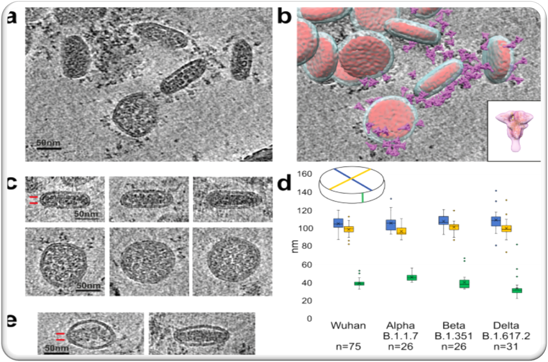
The primary goal was to investigate the three-dimensional architecture of a structure that is assumed to be a virus. The authors performed electron cryotomography (cryo-ET) on suspected SARS-CoV-2 virions and three variants, revealing particles with regular cylindrical morphology. Brief information on cryo-electron tomography: [4]
- First, the object is cooled down up to 4° Kelvin by the use of liquid nitrogen or helium. The lightning-fast freezing preserves the spatial structure of all cell components. At the same time, this method ensures that the cells in the subsequent examination do not burst in the vacuum of an electron microscope. A vacuum is required because otherwise electron beams would be deflected by gas molecules or dust particles.
- After fixation of the sample in the electron microscope, a high-resolution camera produces two-dimensional images of the object from many different tilt angles of the sample.
- As many individual images as possible are required for the subsequent representation of the cell structures in three-dimensional space. At the same time, however, this increases the irradiation time. In order to prevent melting or crystallization of the ice, the lowest possible illuminance levels are used. Computer-aided control of these parameters is intended to prevent spoilage of the sample before completion of the experiments.
- With the help of special computer software, the images obtained are later converted into a three-dimensional image.
Summary electron cryotomography: points of criticism and items to consider
- Irradiation of the sample material can influence the structure.
- Spoilage of the sample material cannot be completely ruled out.
- Three-dimensional images are calculated using an algorithm.
- The images of the publication Calder et al., which were created with the help of cryo-EM, show a cross-section through a cell. One can see cross-sections of typical eversions called microvilli, which the cells use to increase their surface area. In cells that are contained in test tubes, the arrangement of the microvilli is not observed with the same regularity as it is present in cells in the organism, especially if they have previously been damaged and killed during the experiment. As is right and proper, the scientist who made this cross-section of a cell should also have published the adjacent cutting planes. Because then every observer would have been able to recognize that these "circles" are not detached, independent spatial particles, but merely cell processes. But more on that later.
Calder et al. (2022) Methodology
Four different "virus strains" were made available to the authors of the publication:
- Wuhan (hCoV-19/England/02/2020), (GISAID EpiCovTM accession EPI_ISL_407073) from The Respiratory Virus Unit, Public Health England (PHE), UK.
- Alpha variant B.1.1.7 (hCoV-19/England/204690005/2020) from PHE through Professor Wendy Barclay, Imperial College, London, UK.
- Beta variant B.1.351 (501Y.V2.HV001) from the African Health Institute, Durban, South Africa.
- Delta variant B.1.617.2 (GISAID accession EPI_ISL1731019) from Professor Wendy Barclay, through Genotype-to-Phenotype National Virology Consortium.
All "viruses" were grown at the World Influenza Centre, Francis-Crick-Institute, London, UK, under biosafety level 3 conditions in Vero V1 cells provided by Professor Steve Goodbourne, St. George's Hospital, University of London, UK, and stored in Dulbecco's Modified Eagle Medium (DMEM) GibcoTM 41965039 containing 100 U/ml penicillin, 100 μg/ml streptomycin (pen streptococci) and 10% (v/v) heat-inactivated fetal calf serum (FCS).
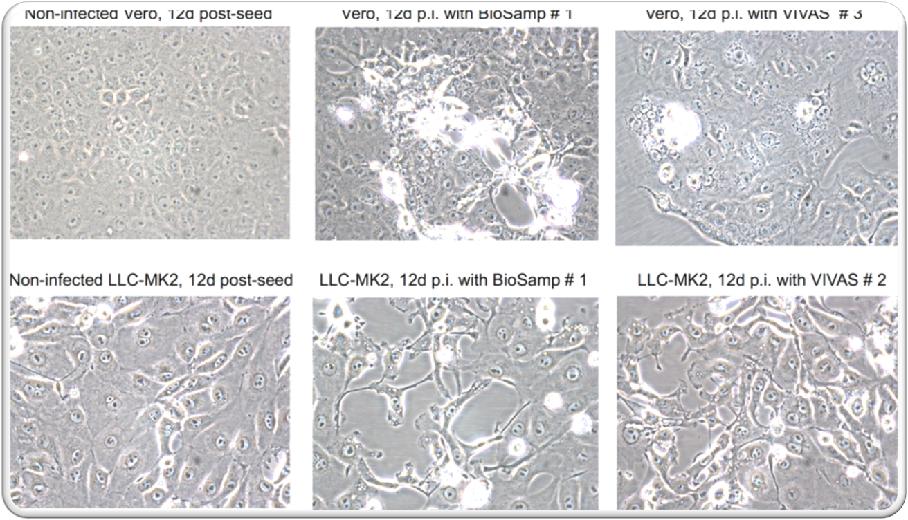
Vero V1 cells were infected with viral semen material at a low infection diversity (MOI) of 0.0001PFU per cell and incubated for 4-5 days as described above until approximately 90% cytopathic effect (CPE) was observed before the supernatant was harvested.
Comments on the methodology during the alleged “infection” and the omitted control experiments
The "viral" samples that were made available to the authors are by no means purified "viral" structures (separated from all other components), but specimens obtained by so-called "isolation in cell cultures", which were created by the occurrence of a so-called cytopathic effect (CPE), which serves in virology as indirect detection of a pathogenic virus. In doing so, a mixture of DNA/RNA from a human and/or from a monkey, a bacterium or a cow is produced, with admixtures of foreign substances and toxic substances. Ergo a mixture of many different substances and genetic material.

What is a cytopathic effect and how can it be recognized?
Virologists kill tissue in the laboratory unnoticed
Virologists don't use the word "isolation" in the proper sense of the word isolation and get suspiciously nervous when asked about it. They understand "isolation" as the production of an effect in the laboratory, the so-called cytopathic effect (CPE), which they also call
a) Infection
b) Proof of the presence of a virus
c) Proof of its multiplication
d) Proof of the destructive power of the assumed virus.
In reality, they kill tissues and cells in the laboratory unnoticed and unwittingly – by starvation and poisoning. This circumstance causes a morphological change in the supposedly "infected" cells.
The alleged cultivation of the virus
This confluence (see picture above) is referred to as giant cell formation and "cytopathic effect". This result of many violent and insane steps (in vitro) is interpreted as the main evidence of the "presence, isolation, multiplication, etc." of the alleged virus. All involved then claim that they have succeeded in cultivating the virus.
The section "Methodology" of the publication Calder et al. (2022) details which method was used in the experimental setup:
The Vero-V1 cells were treated with the following substances:
- Dulbecco's Modified Eagle Medium (DMEM) – Dulbecco's Modified Eagle's Medium, or DMEM for short, is a standardized culture medium for cell culture with broad applicability for human and various animal cells
- 100 U/ml penicillin (a group of antibiotic substances)
- 100 μg/ml streptomycin (Pen streptococci), streptomycin is an antibiotic produced by streptomycete bacteria
- 10% (v/v) heat-inactivated fetal calf serum (FCS) – fetal calf serum is blood serum derived from unborn calves. It is often found in cell culture as a component of culture media.
The added substances alone are problematic and can cause the CPE!
Virological publications:
On December 10, 1954, the idea that the cytopathic effect was a detection method for pathogenic viruses became manifest.
But it was John F. Enders himself who refuted his own assumption at the same time, as he reports that the same death of tissues in the test tube also happens without the addition of supposedly infected material. He expressly warns that the presumption that the presence of a virus could be proven by this effect would have to be thoroughly researched and investigated in the future.
In June 1954, Nobel laureate John Franklin Enders and his colleague Thomas Chalmers Peebles published in the journal "Proceedings of the Society of Experimental Biology and Medicine" No. 86(2), pages 277–286, a report on their work with the alleged measles virus under the title "Propagation in tissue cultures of cytopathogenic agents from patients with measles". [6] As described on page 278 of this publication at the bottom left, the authors used, among other things, the antibiotic streptomycin to “sterilize” throat swabs taken from measles patients, before the cells in the test tube were "infected" with the alleged measles viruses.

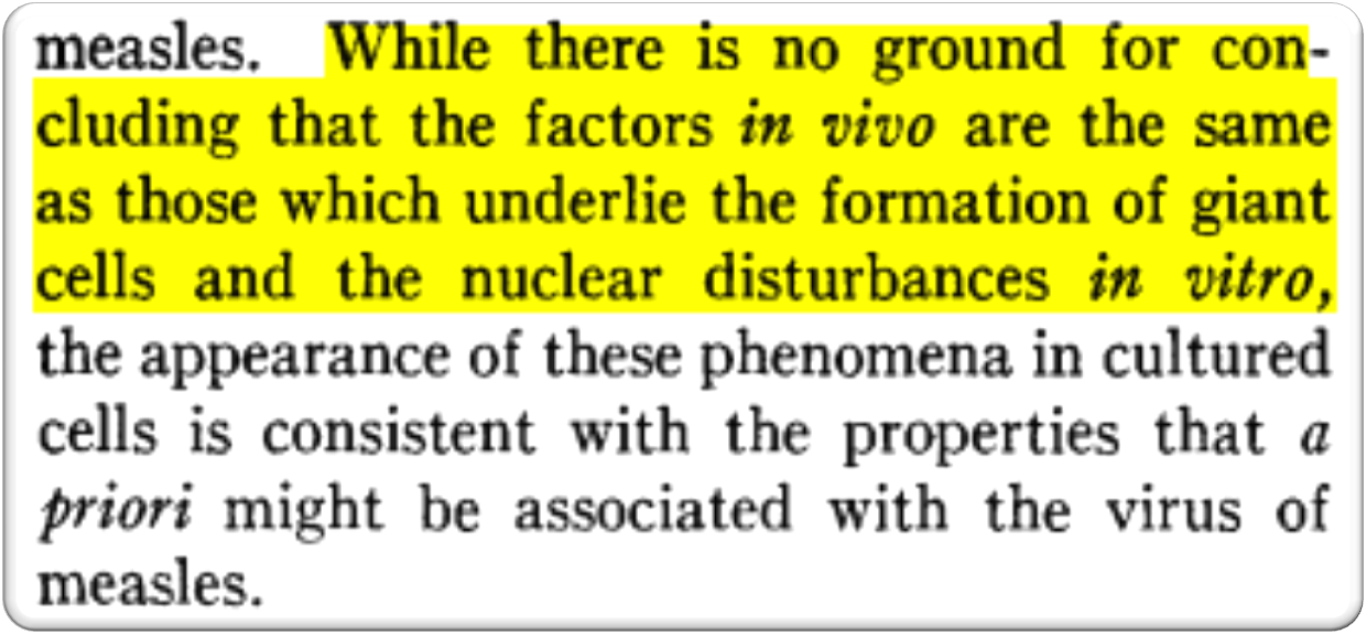
Although the authors of this study pointed out several times (on page 283, left column in the middle and on page 285, right column three times) that the death of the cells is also caused by unknown factors and unknown viruses,

they also claimed two years later that their work from 1954 was fundamental to the production of all future measles vaccines. Despite all the weaknesses and refutations, this study is described by all measles virus supporters as the fundamental study with which the isolation and multiplication of the measles virus has succeeded. This publication is also worth reading for another reason: the authors admit on page 286 that there is no reason to believe that their observations from the test tube have anything to do with those changes in humans that are defined as measles. And it has remained that way to this day.
In 1958 (four years later), the two well-known virologists Beech, V. & von Magnus, P. confirmed in their publication “Studies on measles virus in monkey kidney tissue cultures. [7] Acta Pathologica Microbiologica Scandinavica 42(1):75-85 the speculation that the cytopathic effect is not measles-specific but is caused by other factors.
On page 80 of the publication the following is stated:
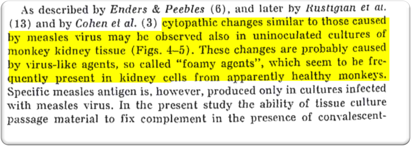
The remarkable thing about this sentence is that it points out the non-specificity of precisely those pathological changes that serve as the starting point for the optical proof of an infection in virology.
Professional Journals:
Science has long known that antibiotics harm mitochondria!
It is common knowledge that streptomycin (an antibiotic used in the treatment of cell cultures before vaccinating an alleged "virus") damages and kills cells [8] [9] [10] by killing the vital bacteria within the cells – the mitochondria, our energy power plants that metabolize oxygen, among other things.
Companies:
The fact that starvation and poisoning of tissues and cells lead to morphological changes, such as the so-called cytopathic effect (CPE) among others, is also known to companies specializing in cell cultures.
According to PromoCell – Human Centered Science: [11]
Antibiotics in cell culture: friend or foe?
The company InCelligence writes: [12]
Antibiotics in cell culture
Self-financed control experiments in various laboratories:
2016: the commissioned laboratory summarized the results as follows: [13]
2021: the commissioned laboratory summarized the results as follows: [13]
Another control within the test series was the addition of so-called yeast RNA (yRNA) to the cell culture.
More about yeast-RNA in the control experiments:
For what reason?
To trigger an SOS reaction analogously in cell cultures (which is known with regard to bacteria), with which dying organisms react by producing a variety of different nucleic acids when they are brought into contact with foreign nucleic acids while dying!
The trick worked, because this way we were still presented with the variety of sequences – without detecting yeast-specific sequences – with which we could calculate 98% of the genome, but without a high number of PCR cycles. In contrast, virologists need two successive PCR steps to obtain the same results, whereby 10-20% of the desired sequences are inserted in the form of synthetic primer nucleic acid pieces to achieve 100%.
The tissues in vitro produce more random sequences in response to the addition of foreign nucleic acids, and these sequences produce in vitro an increased PCR positivity in virology.
Historically, and presently, it is very well documented that the so-called cytopathic effect (CPE) can be caused by various other/external causes that are unrelated to an alleged virus.
Professional Journal Nature & Wikipedia
Another fact that can be added is that there is scientific knowledge that the addition of antibiotics creates "exosomes" (RNA sequences) that were not present before. (Wikipedia 26.10.2020). [14]
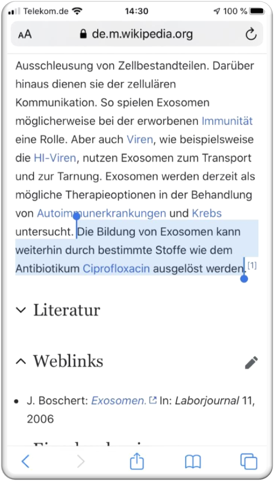
This is among others the result of the study "Antibiotic-induced release of small extracellular vesicles (exosomes) with surface-associated DNA" in the journal Nature. [15]
Consequently, the problem is obvious: according to scientists, exosomes cannot be distinguished from alleged viruses. [16] The most important aspect: exosomes are not specific!
The theory of exosomes serves only as an auxiliary hypothesis and why it is misleading in the resume.
Within the cell theory (which has been refuted) the decay of isolated tissue chunks, which are (mis)interpreted as cells, but also excretions of humans/animals containing connective tissue are interpreted as "exosomes".
The disadvantage of this theory is that both the participants and the recipients, i.e. you, believe the information that virologists have apparently succeeded in finding something specific that implies something (disease, excretion, 5G, etc.).
This also distracts from the fact that no specific RNA or other molecules were detected at all, but only mentally assembled into something. From 1952 onwards, the phages were the inspiration for this, which are the supposed viruses of the bacteria.
More specifically:
The discussion about these structures, which are claimed to be viruses, but also claimed to be harmless but specific structures (exosomes), is completely unfounded.
Because DNA/RNA is constantly changing.
The fictitious construction of genome strands through methods such as sequence alignment and sequence assembly (stringing together many short RNA sequences), does not reflect reality in any way.
Harold Hillman & Gilbert N. Ling - The artifacts of the EM images & refutation of the cell theory
Since this is a rather extensive topic, I will only touch on it briefly to broaden the horizon, otherwise it would go too far beyond the scope of the article.
Discovered decades ago – initially in the heart muscle – and later repeatedly confirmed due to increasingly advanced observation techniques (with which one has to manipulate the tissue to be viewed less and less), strictly speaking, it should be clear to all participants that tissue concentrations (which are intensively interconnected through their gel-like consistency) can only be called cells with disregard of logic and terminology.
Since 1978, a group of scientists connected to the neurologist Harold Hillmann has continuously researched and published the facts in scientific journals, at congresses and in the form of books, why and how the changes of dying, mechanical dissection, chemical fixation and staining of tissue – in order to be able to make it visible in the microscope – make the invisible, gel-like tissue look like as if it were organized in definable units.
In addition, Harold Hillmann and his colleagues have recognized, proven and published extensively that the EM-supported image of cells created by Virchow is wrong and that EM technology has contributed significantly to transporting and stabilizing this image. "Gilbert N. Ling – the liquid in the "cells" is not water."
The summary of the EM recordings:
The only important message to the outside world is this: only "artifacts" are depicted – and the crucial facts are:
- that these images originate only from cell cultures, i.e. dying tissue in a test tube, and definitely do not show anything that is present in a human being,
- that these structures have never been biochemically characterized (sic!),
- nucleic acids have never been obtained from these structures, which are supposed to be the core of the virus (i.e. from a specific structure masqueraded as a virus, the complete nucleic acid has never been obtained).
A motionless image from electron microscopy does not reflect any living biological process. What one is able to observe under the EMs has absolutely nothing to do with what happens in the living organism of man. A result from the laboratory (in vitro) does not allow conclusions to be drawn about dynamic processes within a vital body.
The logical conclusion from the realization that the cytopathic effect was wrongly defined as virus-specific, is and remains the mandatory completion of the control experiments!
The fact that such substances, in combination with the above-mentioned experimental setup – which were also used in the study cited by Peter McCullough – to prepare the cell culture, significantly influence the same and lead to the cytopathic effect, is a fact that has been well documented throughout history. Without control experiments, it cannot be ruled out that the morphological changes of the substrate and the generation of newly occurring gene sequences are causally associated with the poisoning and starvation of the cells.
This knowledge alone in conjunction with laws of thought and logic should absolutely lead to the necessary control experiment – in order to consistently check one's own results – with all combinations of the experimental setup and also the logging, to verify whether this effect also occurs when an uninfected material is added to the cell culture to be infected (e.g. yeast RNA yRNA), or when the cell culture is pretreated in exactly the same way as if one intended to "infect" it in order to rule out that the experimental setup itself is not the cause of the result. These control experiments were not and are not performed.
In the publication mentioned by Peter McCullough and Co. [1], control experiments are not documented, not even to some extent.
Virus growth and purification for EM Calder et al. (2022)
With regard to cleaning before taking EM images the publication states:
Since most readers probably feel overwhelmed with these technical terms, I will summarize the most important key messages for you.
Prior to the purification step, within the starved, poisoned and inoculated cell culture there are numerous host cell organelles and other proteins, microorganisms, bacteria, a mixture of RNA, produced from a human and/or a monkey, bacteria or a cow with admixtures of foreign substances and pollutants and much more.
The supernatant of all these structures and the genetic mixture that occurs during the morphological change (called CPE) is then subjected to preliminary cleaning through further steps.
Based on the documentation of the publication Calder et al. (2022) it becomes obvious that no clean isolation was performed. As the authors themselves describe, only an intermediate step of purification was inserted, in the form of sedimentation. It was pelletized.
Simple pelletizing is usually an early step as part of the complex and multi-stage processes for purifying particles. The supernatant or pellet is then discarded or further processed. The more careful cleaning, on the other hand, would be density gradient centrifugation. [17]
Density gradient centrifugation is used, for example, for further purification and separation of particle fractions from differential centrifugation. In contrast to this method, a solvent with a density gradient is used here. As a result, fractions can be better separated from macromolecules. A substance (e.g. sucrose) is added to the solvent, the concentration of which varies relative to the distance to the axis of rotation. In the sample tube, this results in a location-dependent, varying density, a density gradient that acts on the macromolecules to be fractionated during the centrifugation run. This results in clearly separated bands with fractions of the particles, which can be used for further preparative or analytical purposes. [17]
Ultracentrifugation in the application of sedimentation, as used in this publication, only allows the enrichment of the samples, but no clean purification.
Specifically: pelletizing is the concentration of solid components by centrifugation from the liquid to the bottom of the centrifuge glass. Here, everything gathers that cannot remain soluble; nothing is therefore isolated. The pellet, consisting of proteins and fats, is transformed together with the detergents/solvents into soap bubbles (= micelle) by swirling (through suction and expulsion), then mixed with dyes, dried and labelled as viruses in the EM.
This type of "cleaning technique" makes it impossible for the authors to obtain the exact density and determine the degree of purification of an EM image.
This type of purification (sedimentation/pelleting), as used in this publication by Calder et al. (2022), was also the method used in one of the two globally prevailing publications on SARS-CoV-2 from China, [19] which were used as a template for all other publications worldwide.
Na Zhu & Wenjie Tan et al. correspondence confirm our statements
This approach does not even seem to be known by many scientists, among them Prof. Kämmerer or Prof. Bhakdi.
Prof. Kämmerer stated in the CA (Corona Ausschuss – Corona Investigative Committee) session #22 - section 03:32:05 - 03:35:30 [18] that in the publication "A Novel Coronavirus from Patients with Pneumonia in China, 2019" by Na Zhu & Wenjie Tan et al. [19] clearly a virus was carefully isolated.
In order to show that this type of cleaning definitely does not produce clean isolation, and also does not make it possible to say how high the density is or that it is suitable for determining the degree of purification of the EM images, the authors of the publications were directly contacted.
The authors of the publication refute Prof. Kämmerer's statement in writing and confirmed our statements that:

Wan Beom Park et al. correspondence confirm our statements
Prof. Sucharit Bhakdi & Prof. Karina Reiß claimed in a correspondence with us [21] that a clean isolation and thus the proof of the existence of SARS-CoV-2 had been provided in the publication "Virus Isolation from the First Patient with SARS-CoV-2 in Korea" by Wan Beom Park et al. [22]
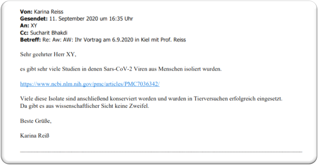
When we asked the authors of the publication directly whether the electron micrographs presented in their in vitro experiments depict purified viruses, one of the authors himself confirmed in writing, and thus automatically refutes the statements of Prof. Bhakdi and Prof. Reiß:
On March 19, 2020, Wan Beom Park et al. replied:
➥ Continue reading Part 2 here
➥ List of References