Peter McCullough interprets a cell’s own structures as "viruses" 2
NEXT LEVEL - Wissen neu gedacht
Article part 1 <----------------------
On March 19, 2020, Wan Beom Park et al. replied:
These examples illustrate that even well-established scientists in this very field, even those who have gained an enormous reputation over decades and have themselves trained hundreds of scientists, either do not possess or overlook the knowledge of such subtleties and thus automatically make incorrect predictions and statements.
Because many laymen trust these authorities and are not even able to read or understand these publications, they pass on erroneous information to their fellow human beings.
Just as it happened in this case with Peter McCullough and the many educational channels such as ScienceFiles, Dr. Bodo Schiffmann, #WirMachenAuf and others.
Based solely on inadequate research and blind trust, progress is thus actively prevented, and people are irritated and misguided. These misinterpretations have serious consequences because they provide the illusion that a viral structure has actually been detected. The fear in the population is maintained, which leads to compromised health for some and which further legitimizes the irrational actions of politicians and their stooges.
Summarized in simple terms:
It is important to understand that this publication Calder et al. (2022), which is referenced by Peter McCullough and all copiers such as ScienceFiles, Dr. Bodo Schiffmann and others, only talks about pelletized (sedimented) matter and not isolated by a sucrose density gradient (density gradient centrifugation) AND that the pelletized matter at no point in time was ever biochemically characterized. Furthermore, genome sequencing was also missing to determine whether the structures shown carry any gene sequences attributed to SARS-CoV-2.
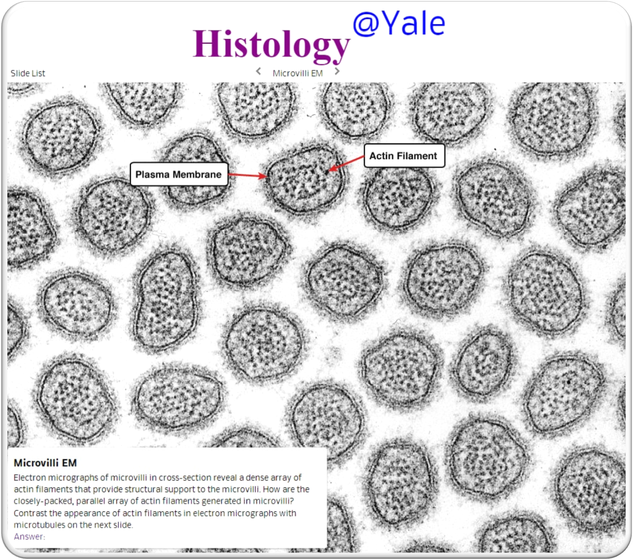
Even during the step of taking the EM images, no control experiments are documented in the aforementioned publication at all in order to rule out that these structures are not the cell's own material (e.g. microvilli in cross-section).
The minimum requirement for controls would have been:
- To document a control experiment with which it can be completely ruled out that the cytopathic effect – which is claimed by virologists as proof of the existence of a virus – is caused by the experimental setup itself.
- To document a control experiment whether the same structures can be found under the electron microscope with the same treated cell culture and the addition of different substances, but which have not been inoculated with an allegedly infected material.
- The same test setup to investigate and document whether identical results are obtained when "infecting" several common primate and human cell lines, including human adenocarcinoma cells (A549), human liver cells (HUH 7.0) and human embryonic kidney cells (HEK-293T), in order to rule out that the cell line used, in this case Vero V1, is not the cause.
What are these structures that are recognizable on the EM images and are falsely passed off as viruses?
Crucial things must be said in general about the EM images:
Structures shown in EM images and published as images of viruses have never been biochemically characterized. No nucleic acid has ever been taken from such particles and determined. These particles are only passed off as viruses and at the same time one important fact is withheld; namely that the same particles of this type are also formed every time "uninfected" cell cultures are treated in the same way as those cell cultures defined as "infected". Non-virologists refer to these particles as phagosomes, endosomes, exosomes, transport vesicles and in cross-section as villi etc. pp.
You will find the same pictorial representation for many different claimed structures.
EM images always show only dead, chemically fixed things. [23] The figure shows soap micelles made from detergents, fats and proteins that have been preserved by freezing and may have been formed by this freezing process to begin with.
Again, the following figure shows identical structures, just like they are published with regard to SARS-CoV-2 or other viruses, in the form of liposomes. The term liposome is composed of the Greek terms "lipos" for fat and "soma" for body. Liposomes consist of one or more lipid bilayers that are deposited around an aqueous core. The size of the microscopically small vesicles is subject to a fluctuation range of 50 to 1000 nm.
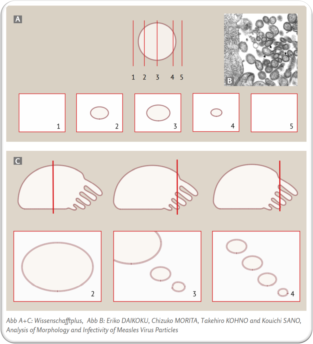
The circle and the cylindrical structures shown in EM images
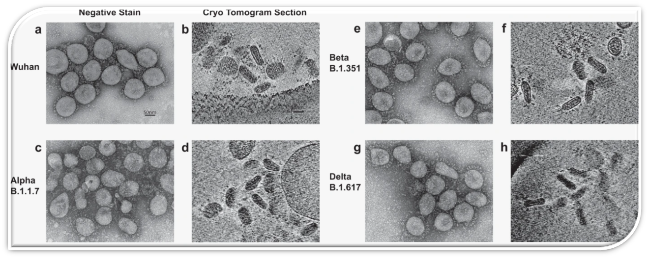
The images taken by the cryo-EM in the publication Calder et al. 2022 [1] show a cross-section through a cell. You can see cross-sections of typical protuberances, called microvilli, with the help of which the cells increase their surface area. In cells in the test tube, the arrangement of the microvilli is not as uniform as in the cells in the organism, especially if they have previously been experimentally damaged and killed. If the scientist who took this cross-section image of a cell had worked correctly, he would also have had to publish the cutting planes above and below this cutting plane. But then every observer would have been able to notice that the circular structures are not detached, spatial particles, but instead a cross-section of cell extensions (round, amoeba-like extensions).
Because the cells studied came from supposedly "infected" cells, the authors claim ad hoc that the cross-sections through the villi are cross-sections of SARS, SARS-CoV-2, measles and other viruses. In an extremely unscientific manner, they suppress the cutting planes in front of and behind the image shown (Figure 15 illustrates these planes). Only the documentation of these additional cutting planes could have shown whether the cross-sections shown were cell protuberances or independent particles. On the basis of such incompletely investigated cell-specific structures, the authors determine the diameter of the suspected viruses.
Even if it had been shown that identifiable independent particles occur in the cross-sections, this would only have demonstrated the presence of typical cell-own transport particles, which are called, for example, "exosomes". In order to prove that separate particles, which have never been proven in this publication and in other SARS and measles studies, are viruses, they would have had to be isolated, photographed and biochemically characterized. To this day, this has not been done for the SARS-CoV-2 or any other pathogenic virus and has not been documented in any study.
To claim that the cells used would produce SARS-CoV-2 viruses, the authors pelletize membrane components, nucleic acids and proteins of dead cells by centrifugation. The images of the pellets clearly show their composition from cell fragments. An incomplete intake of a pellet is inadmissibly referred to as "SARS-CoV-2 particles", although its composition has not been biochemically determined in this publication or in any other studies.
Control experiments in which cell fragments of dead but not "infected" cells are compressed into pellets and compared with the pellets from "infected" cells were not performed.
Therefore, these experiments do not allow any other statement, except: that the cell's own components were used!
The authors of this publication did not conduct or document any control experiments at all.
In other studies, normally treated cells not prepared for "infection" are used as control specimen. But these untreated cells are not observed and included in the experiments in the same manner, rather only in a – literally: "similar" manner – and beyond that in an undescribed and undocumented way, and actually not at all, as the review of the publications shows.
If you perform a Google search for the term "Cross section microvilli EM," you will find many images of cross-sections through microvilli and realize that these structures, which are mistakenly interpreted as viruses, occur everywhere.
The only important message to the outside world is this: only "artifacts" are depicted – and the crucial facts are:
- that these images originate only from cell cultures, i.e. dying tissue in a test tube, and definitely do not show anything that is present in a human being,
- that these structures have never been biochemically characterized (sic!),
- nucleic acids have never been obtained from these structures, which are supposed to be the core of the virus (i.e. from a specific structure masqueraded as a virus, the complete nucleic acid has never been obtained).
An example of biochemical characterization can be found in one of the publications of Dr. Stefan Lanka: in his work on a so-called giant virus [25] (actually bionts).
At first glance, the term "virus" in Dr. Stefan Lanka's work seems contradictory when looked at from a superficial point of view. In fact, however, it is not.
In 1992, in the City of Amsterdam, Dr. Stefan Lanka met Eleni Papadopulus-Eleopulus from the Perth Group – this developed into a collaboration and a personal friendship.
She assured Dr. Stefan Lanka that not only is there no HIV, but that it applies to all "viruses".
By the year 1998, Dr. Lanka had finalized his personal review of all virus types, resulting in this apparent contradiction.
When Dr. Lanka published his virology paper in 1993 [25], Eleni Papadopulus-Eleopulus was irritated, whereupon Dr. Lanka explained to her in one of the many telephone calls that he had to grudgingly accept that the professors who revised his version removed the "symbionts" and the explanations about them and replaced them with "virus".
In the case of algae (Ectocarpus siliculosus), he describes this step on p. 507 and p. 509.
Here you can see the extracted ring-shaped and closed DNA strand.
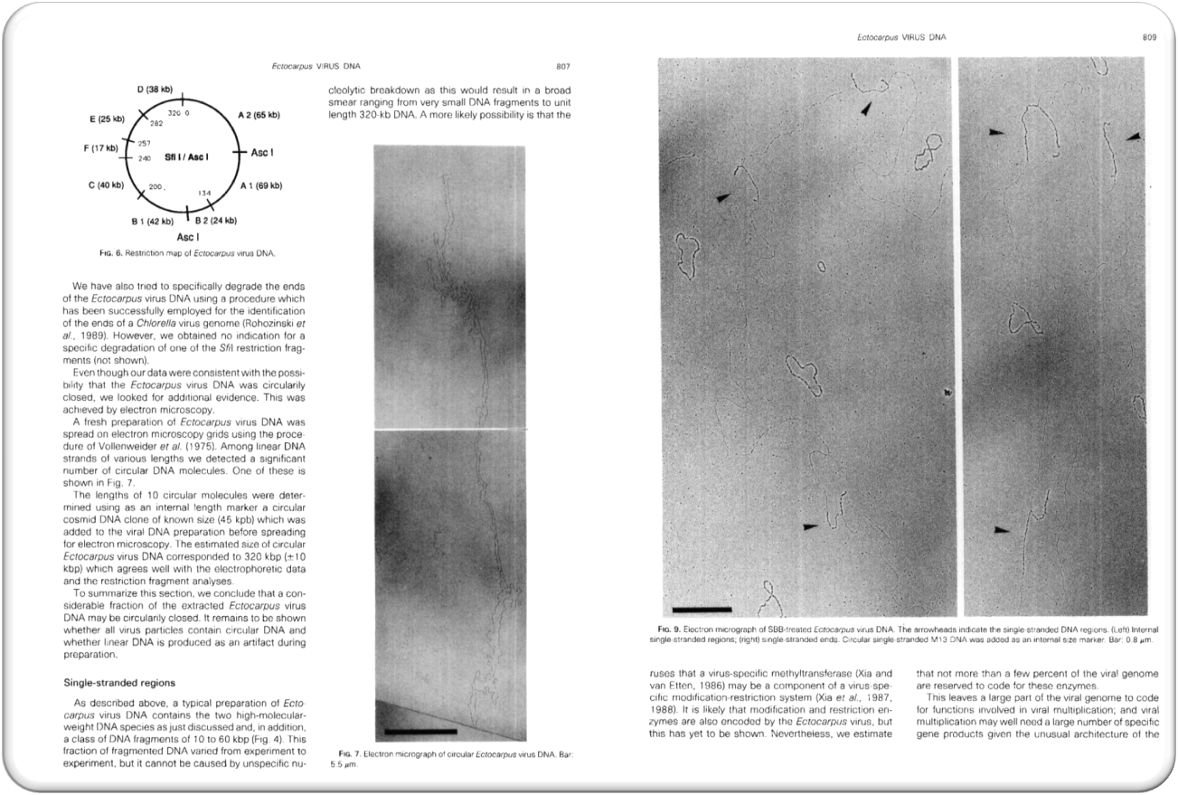
From all types of "phages" and "giant viruses", the nucleic acids of exactly the same length and exactly the same composition can be obtained each time (see figure above). But it has never been possible to isolate a nucleic acid (DNA or RNA) from a structure or fluid whose length and composition would correspond to what virologists claim to be the genetic strand of a pathogenic virus.
Why the virologists have entirely gotten lost in a completely detached and dangerous anti-science, becomes clear from the sequence of events that happened between 1951 and December 10, 1954. After medical virology was deprived of its basis by control experiments in 1951 [26], from 1952 onwards, the phages of bacteria were then established as the model for the persistent ideology of what "disease-causing viruses" should look like:
a nucleic acid of a certain length and composition, surrounded by a shell consisting of a certain number of specific proteins.
But:
- due to a lack of electron micrographs of "pathogenic viruses" in humans/animals/plants,
- due to a lack of biochemical characterization of the components of "pathogenic viruses",
- and due to a lack of isolation,
virologists were and are still forced to assemble individual components from supposedly "virally" diseased tissue mentally and graphically into viruses and to present these mental products to themselves and the public as existing viruses under false pretenses!!
In summary: Electron micrographs, which supposedly show viruses, are known and typical artefacts or cellular structures
Virologists publish a variety of electron micrographs of structures they call viruses. In doing so, they conceal the fact that ALL these images are only typical structures of dying cell cultures or represent protein-fat-soap bubbles produced in the laboratory, and were NEVER photographed in humans/animals/plants or in liquids obtained from them.
Non-virological researchers refer to the same structures, which virologists pass off as viruses, as either typical cell components such as villi (amoeba-like protuberances used by cells to cling to the substrate and move), or "exosomes" or "virus-like particles." Thus, a further, independent proof has been provided that the claims of virologists of being able to see viruses in the electron microscope have been scientifically refuted.
Comparison: SARS-CoV-2, measles virus and cross-sectional images of microvilli [27]
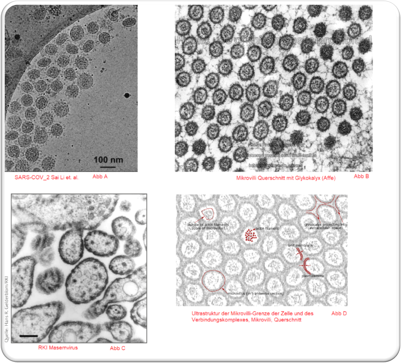
Picture B – Cross-sectional image of microvilli with glycocalyx (monkey)
Picture C – RKI measles virus
Picture D – Ultrastructure of the cell microvillous border and junctional complex, microvilli, transverse section
List of References:
[1] https://www.nature.com/articles/s42003-022-04183-1#Sec13
[2] Peter Mc Cullough: https://www.trialsitenews.com/a/electron-cryotomography-of-sars-cov-2-virions-6fe352f0
[3] ScienceFiles: https://sciencefiles.org/2022/11/19/doch-es-gibt-viren-forscher-machen-nahaufnahme-von-sars-cov-2/
[4] Wikipedia: https://de.wikipedia.org/wiki/Kryoelektronentomographie
[6] Prof. John Franklin Enders Masernpublikation: („Propagation in tissue cultures of cytopathogenic agents from patients with measles“) https://pubmedinfo.files.wordpress.com/2017/01/propagation-in-tissue-cultures-of-cytopathogenic-agents-from-patients-with-measles.pdf
[7] STUDIES ON MEASLES VIRUS IN MONKEY KIDNEY TISSUE CULTURES bech-von-magnus-1959-1.pdf (wordpress.com) |
https://therulingclassobserver.files.wordpress.com/2020/06/bech-von-magnus-1959-1.pdf
[8] https://www.pharmazeutische-zeitung.de/ausgabe-282013/antibiotika-schaden-mitochondrien/
[10] Bactericidal Antibiotics Induce Mitochondrial Dysfunction and Oxidative Damage in Mammalian Cells https://stm.sciencemag.org/content/5/192/192ra85.short
[12] https://incelligence.de/zellkultur/zellkultur-kontaminationen/antibiotika
[13] Kostenlos Verfügbar: https://telegra.ph/Kontrollexperiment-Phase-1---Die-Widerlegung-der-Virologie-durch-den-cytopathsichen-Effekt-03-10
[14] Exosom (Vesikel) https://de.m.wikipedia.org/wiki/Exosom_(Vesikel)
[15] NATURE: "Antibiotic-induced release of small
extracellular vesicles (exosomes) with surface-associated DNA" https://www.nature.com/articles/s41598-017-08392-1
[16] NCBI: Rockefeller Education: When is a virus an exosome?
https://www.ncbi.nlm.nih.gov/pmc/articles/PMC2248418/
[17] https://de.wikipedia.org/wiki/Ultrazentrifuge
[18] CA-Sitzung 22 - Abschnitt 03:32:05 - 03:35:30 https://odysee.com/@DD-Zone:3/Corona-Ausschuss-22_b:6
[19] CCDC: A Novel Coronavirus from Patients with Pneumonia in China, 2019
https://www.nejm.org/doi/full/10.1056/nejmoa2001017
[20] Mail Wenjie Tan: https://t.me/Corona_Fakten_Video_Backup/117
[21] Mail Bhakdi: https://t.me/Corona_Fakten_Video_Backup/111
[22] Mail Wan Boem Park: https://t.me/Corona_Fakten_Video_Backup/118
[23] https://docplayer.org/4939490-Arbeiten-mit-membranproteinen-rupert-abele.html | S.20 und S.26
[24] Querschnitt Mikrovilli: http://medcell.org/histology/epithelia_lab/microvilli_em.php
[25] Genome Structure of a Virus Infecting the Marine Brown Alga Ectocarpus siliculosus
https://www.sciencedirect.com/science/article/abs/pii/S004268228371189X
[26] Prof. Karlheinz Lüdtke, Max-Planck-Institut für Wissenschaftsgeschichte, Frühgeschichte der Virologie, Sonderdruck 125, 89 Seiten, 1999. i. K. (A 2) Preprint 1999.
https://www.mpiwg-berlin.mpg.de/Preprints/P125.PDF
The experiments, however, did not yield any growth of the filterable virus. In hopes of triggering the virulence of a filter-passing virus, animal experiments were also conducted. But the results were negative every single time. It was not possible at all to grow a true filter-passing virus from the filtrate by further inoculation on various culture media.
[27]
Abb A: Molecular Architecture of the SARS-CoV-2 Virus
Molecular Architecture of the SARS-CoV-2 Virus - PMC (nih.gov)
Abb B: Microvilli cross section with glycocalyx (monkey)
http://www.drjastrow.de/WAI/EM/externes/Wartenberg/Mvilli1.jpg
Abb C: RKI: Masernvirus: https://www.rki.de/DE/Content/Infekt/NRZ/EM/Aufnahmen/EM_Tab_Masern.html
Abb D: Ultrastructure of the Cell microvillous border and Junctional Complex, microvilli, transverse section
https://www.bu.edu/phpbin/medlib/histology/p/20602loa.htm