Sperm Cell

🛑 ALL INFORMATION CLICK HERE 👈🏻👈🏻👈🏻
Sperm Cell
Certified Medical Magazine by WMA, ACSA, HON
What’s the Function of a Sperm Cell? – Definition & Structure
What’s the Function of a Sperm Cell? – Definition & Structure
By Antonio Barberá BSc (embryologist), Emilio Gómez Sánchez BSc, PhD (senior embryologist), Zaira Salvador BSc, MSc (embryologist) and Sandra Fernández BA, MA (fertility counselor).
By Emilio Gómez Sánchez BSc, PhD (senior embryologist).
By Zaira Salvador BSc, MSc (embryologist).
By Zaira Salvador BSc, MSc (embryologist).
By Zaira Salvador BSc, MSc (embryologist).
By Zaira Salvador BSc, MSc (embryologist).
By Zaira Salvador BSc, MSc (embryologist).
By Zaira Salvador BSc, MSc (embryologist).
By Zaira Salvador BSc, MSc (embryologist).
By Zaira Salvador BSc, MSc (embryologist).
Bachelor's Degree in Biology and Biochemistry from the University of Valencia (UV). He has developed several research projects for the Research Foundation La Fe (Valencia, Spain). He has been working as a biologist and biochemist at the andrology lab of CREA Valencia for over 8 years. More information about Antonio Barberá
Bachelor's Degree in Biology from the University of Seville. PhD in Biology from the University of Valencia. Large experience as an Embryologist Specialized in Assisted Reproduction. Currently, he is the IVF Lab Director of Tahe Fertilidad. More information about Emilio Gómez Sánchez
Bachelor's Degree in Biotechnology from the Technical University of Valencia (UPV). Biotechnology Degree from the National University of Ireland en Galway (NUIG) and embryologist specializing in Assisted Reproduction, with a Master's Degree in Biotechnology of Human Reproduction from the University of Valencia (UV) and the Valencian Infertility Institute (IVI) More information about Zaira Salvador
Bachelor of Arts in Translation and Interpreting (English, Spanish, Catalan, German) from the University of Valencia (UV) and Heriot-Watt University, Riccarton Campus (Edinburgh, UK). Postgraduate Course in Legal Translation from the University of Valencia. Specialist in Medical Translation, with several years of experience in the field of Assisted Reproduction. More information about Sandra Fernández
What Is The Function of Sperm Cells?
How do you know when a man is fertile?
Treatment : Which one do I need? How much will it cost me? Clinic : Which one do I choose? What aspects should I consider?
Generate report
Male Fertility – Parts & Functions of the Male Reproductive System
Fertility in males depends on the proper functioning of their reproductive system. Learn about its anatomy with pictures and definitions of its organs. Read more
It takes place in testicles and lasts two months and a half approximately. It is the maduration of some cells that split, madurate, and eventually become spermatozoa. Read more
Sperm’s Journey to the Egg – How Sperm Meets Egg with Pictures
How long does it take for sperm to reach the egg? When and where does sperm meet egg? Get the answer to these questions about the sperm's path to the egg. Read more
The information provided on this site is designed to support, not replace, the relationship that exists between a patient/site visitor and his/her existing physician.
Certified by Health Quality Agency of Andalusia
Certified Medical Website by the Official College of Physicians of Barcelona
W3C WAI-AA Web Content Accessibility
Busines Adapter certificate in compliance with the LSSI (Spanish Information Society Services Act)
inviTRA Copyright © 2021. Powered by DCIP Consulting.
We use cookies necessary for navigation and traffic analysis. We do not use cookies that provide us with personal data or advertising cookies.
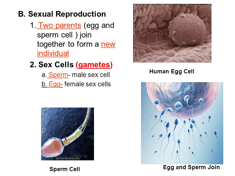
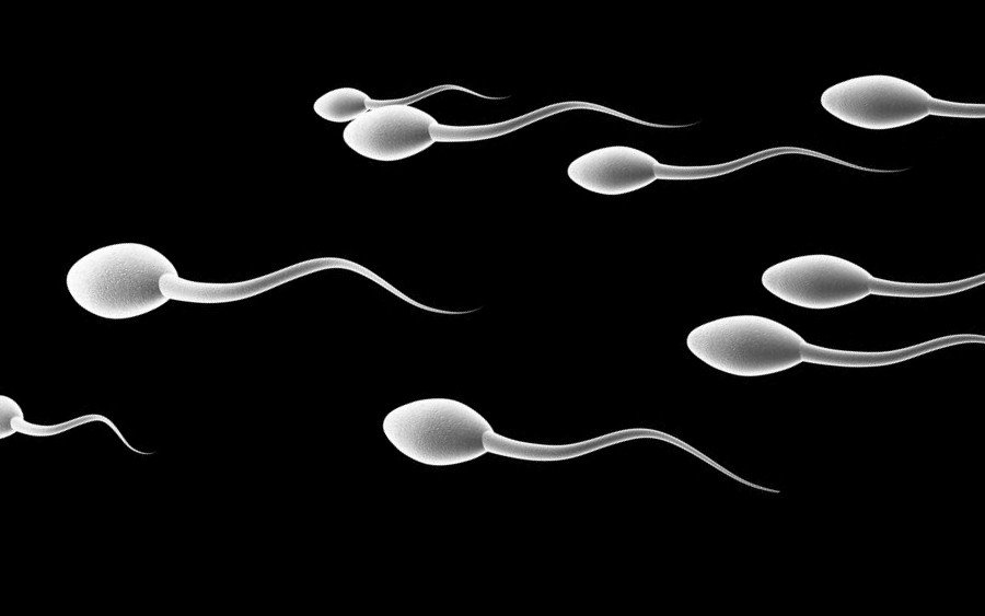
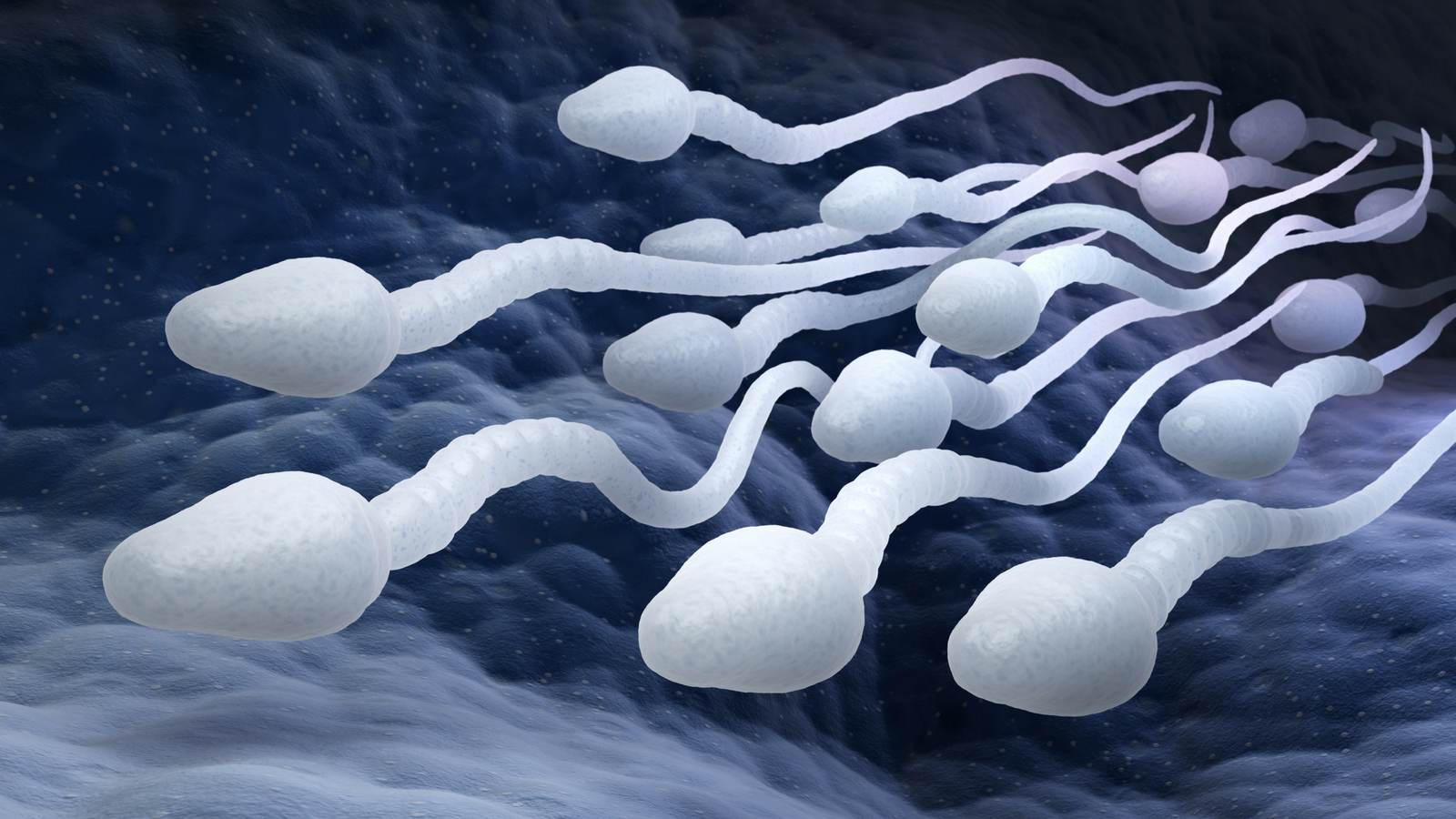
A spermatozoon, in plural spermatozoa , or sperm cell is the male reproductive cell that is expelled along with the seminal fluid or semen when a man ejaculates. In humans, spermatozoa determine the gender of the baby-to-be, which means that they can carry either the X or the Y chromosome.
The function of spermatozoa is to fertilize the egg cell during fertilization, creating a new organism called zygote that will develop from embryo to fetus during the 9 months of pregnancy. Human sperm cells are haploid, which means that they contain 23 chromosomes.
Below you have an index with the 10 points we are going to deal with in this article.
The sperms, spermatozoa or sperm cells are the male sex cells. Their function is to combine with the female sex cell and create a completely new organism. Spermatozoa are expelled with seminal fluid (semen) during ejaculation.
Spermatozoa production takes place in the testis, while oocytes or egg cells are produced in the ovaries of the woman. When an egg and a spermatozoon unite, they create a zygote, which will develop into an embryo (an unborn, developing organism), and later into a fetus.
Learn about the differences between these terms with this post: Differences between human zygote, embryo and fetus .
In humans, the sperm cell is the one responsible for determining the gender of the new organism . This means that spermatozoa can carry either a Y or an X chromosome, while egg cells always contain an X chromosome.
Human sperm cells are always haploid, that is, they contain 23 chromosomes. When a spermatozoon reaches and joins an oocyte, which also contains 23 chromosomes, they form a diploid cell of 46 cells.
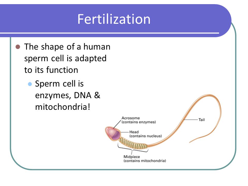
Spermatozoa are structurally specialized to its function through the following anatomical changes:
According to experts, actually spermatozoa have a very narrow function in comparison with other cells that adapt to their function as well. For example, they lack ribosomes, since they are actually unnecessary for carrying DNA to the egg cell.
Before ejaculation, spermatozoa pass through the ejaculatory ducts, and mix with fluids from the seminal vesicles, bulbourethral glands, and the prostate. In fact, 65-75% of the semen expelled contains fluids from the seminal vesicles, including:
Also, the sertoli cells secrete a fluid into the seminiferous tubules that helps in the transport of spermatozoa to the genital ducts. In short, semen is typically translucent with white or grey tint, sometimes yellowish.
The presence of blood in the semen is known as hematospermia and can cause a reddish or pinkish color. If this happens, it may indicate a medical problem that should be evaluated by your doctor.
Very simply put, seminal fluid is comprised of proteins, fructose, water and other components such as vitamins, minerals, etc. So, dehydration, a lack of nutrients, or vitamin deficiency can lead to male infertility if untreated.
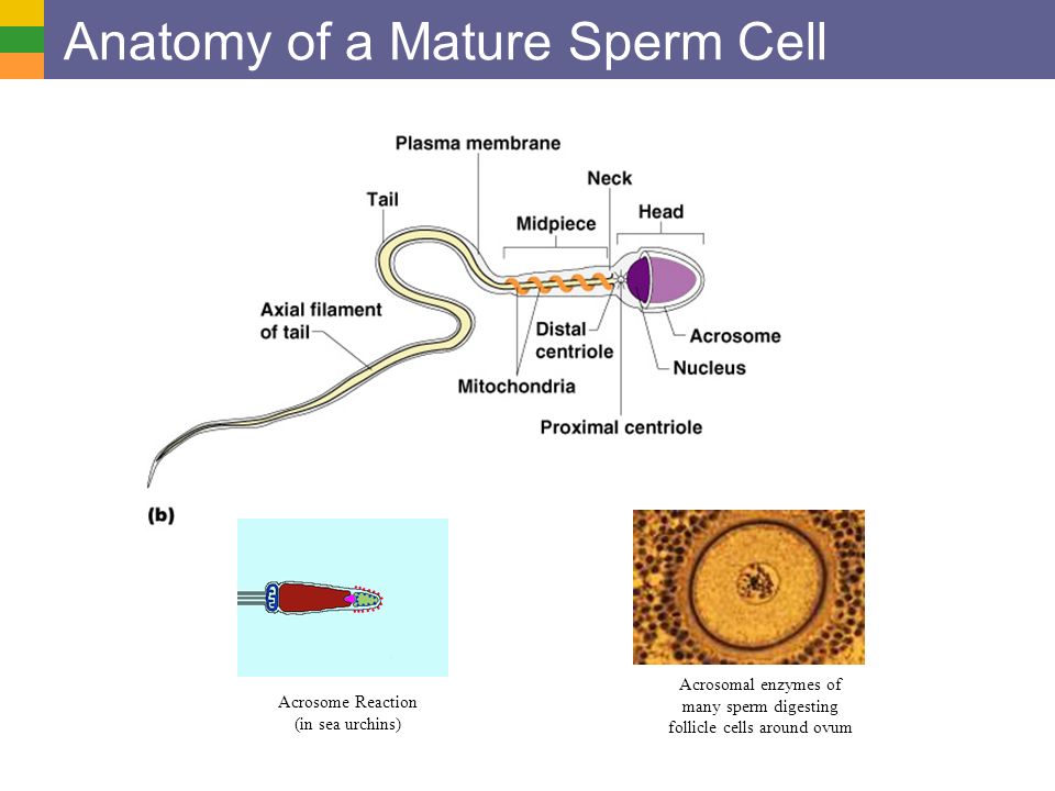
The scientist Antonie van Leeuwenhoek was the first person who described in detail the structure of a sperm cell in 1677. Although the parts of spermatozoa are more or less common in all mammal species—a head and a long tail—there are small differences between species, especially in the morphology of the head.
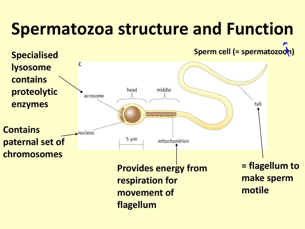
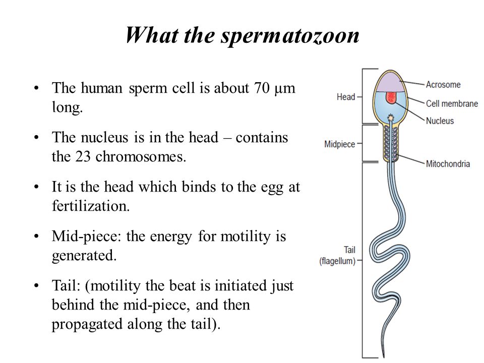
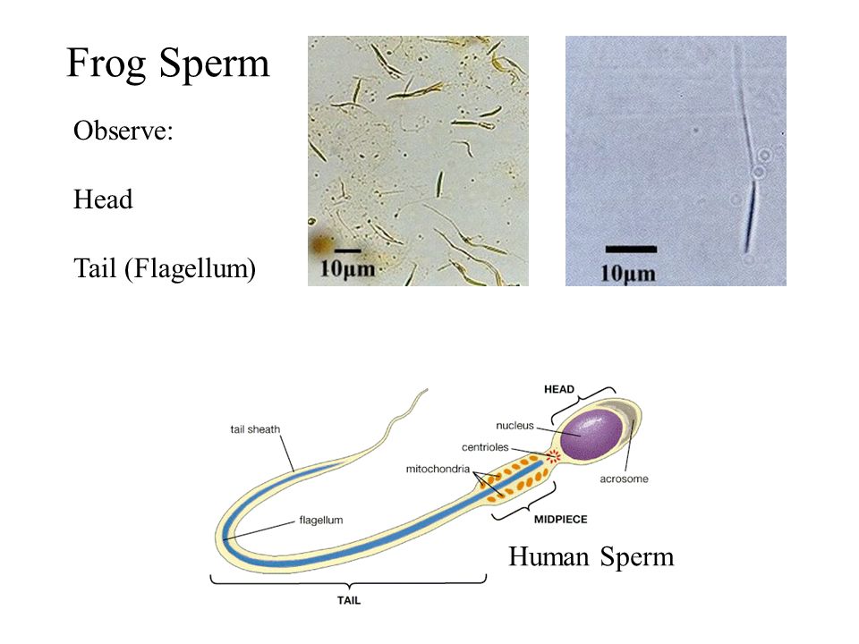
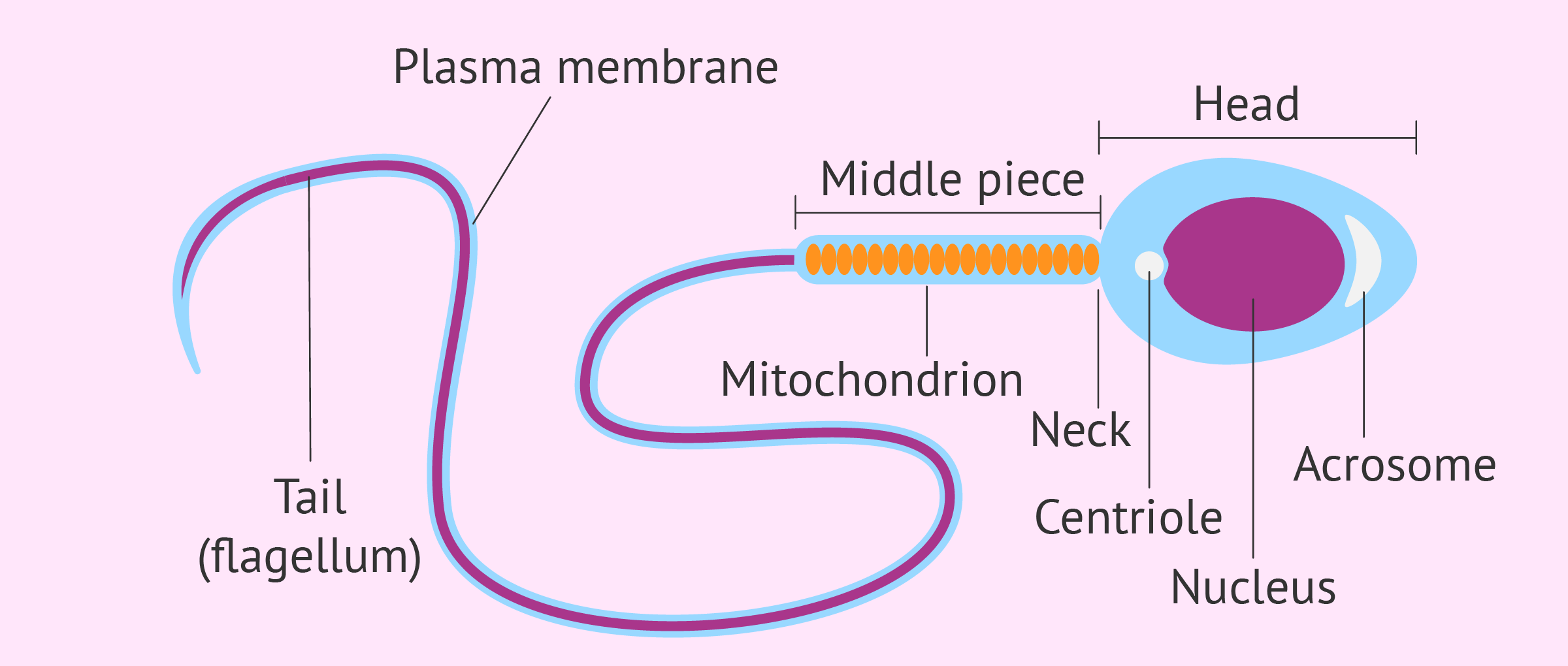
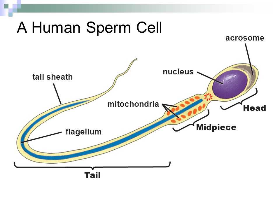
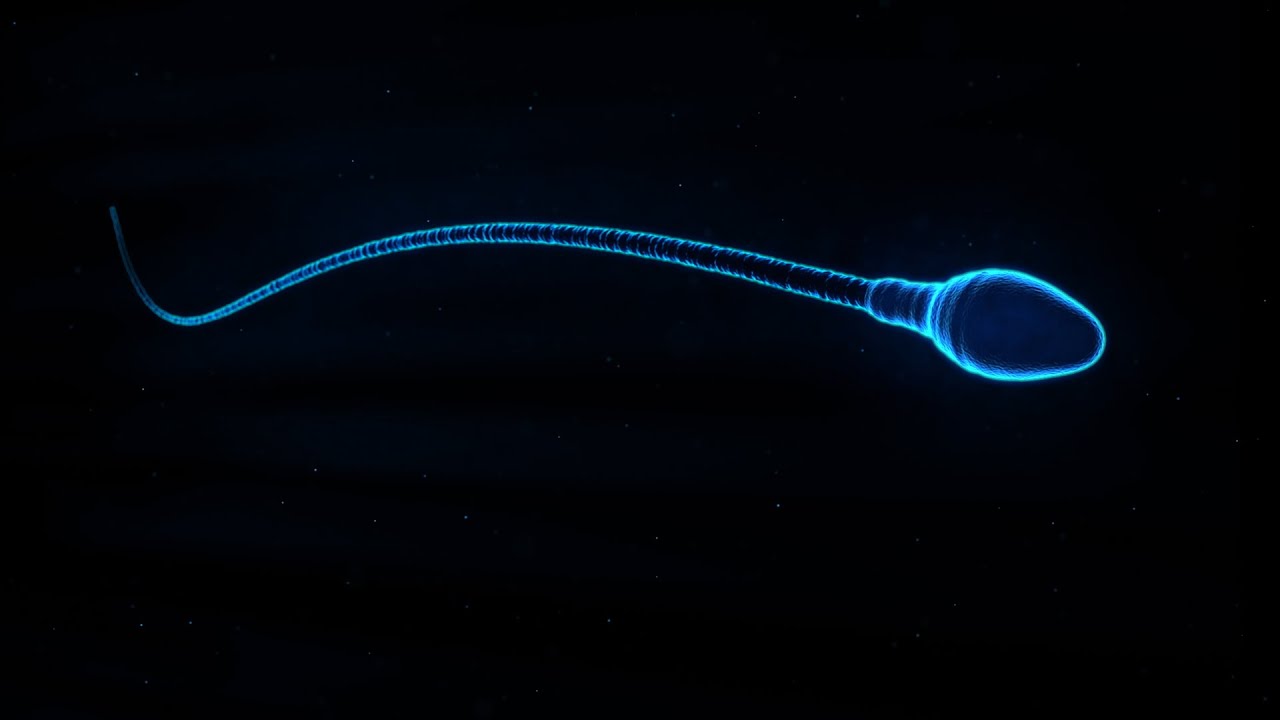
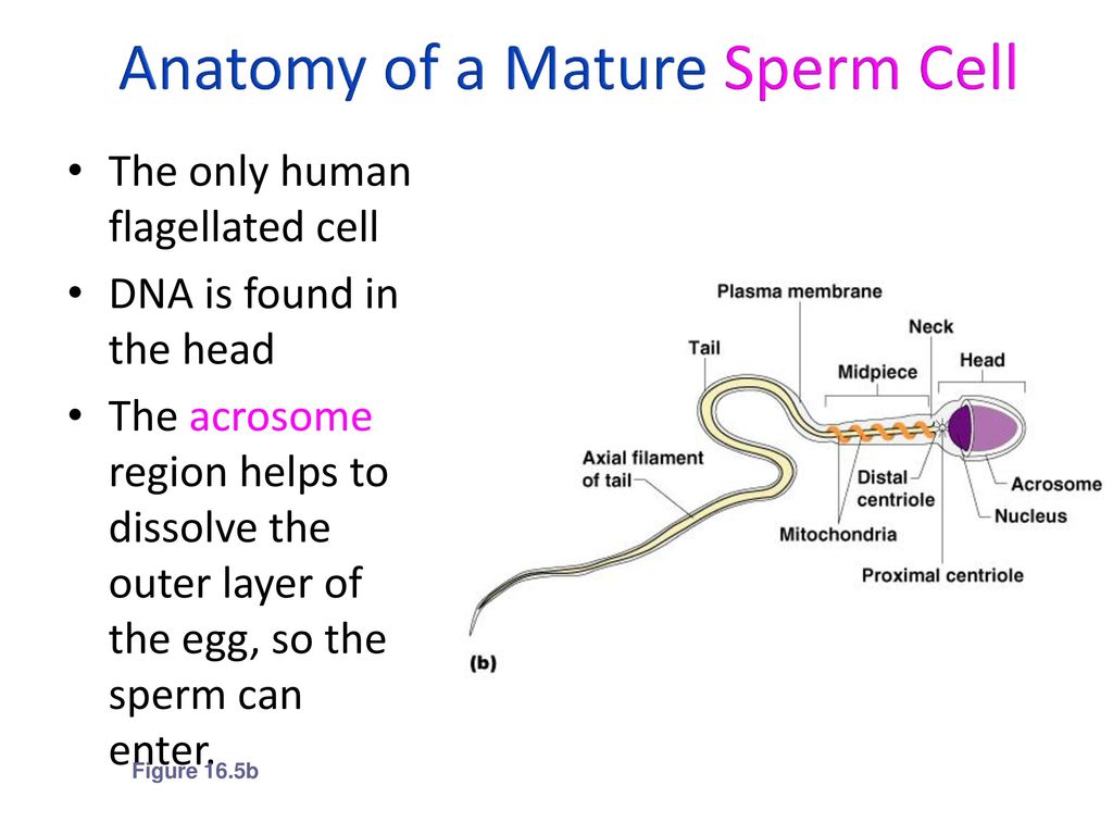
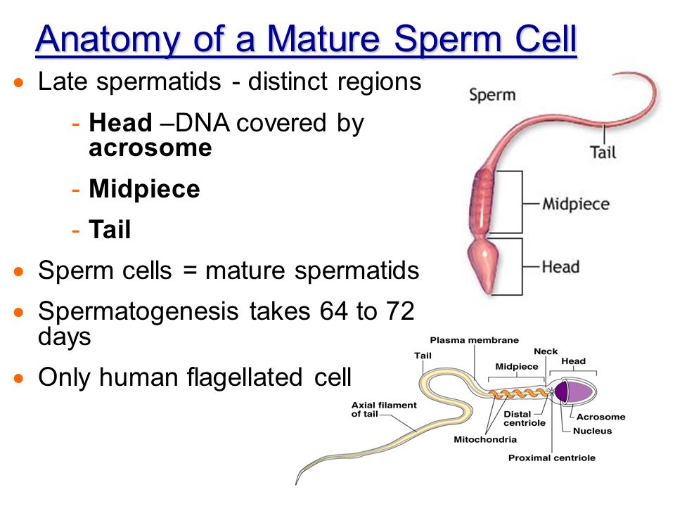
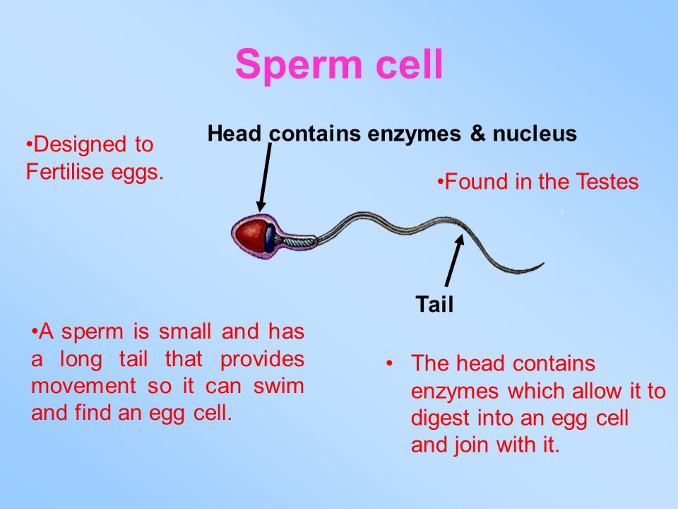
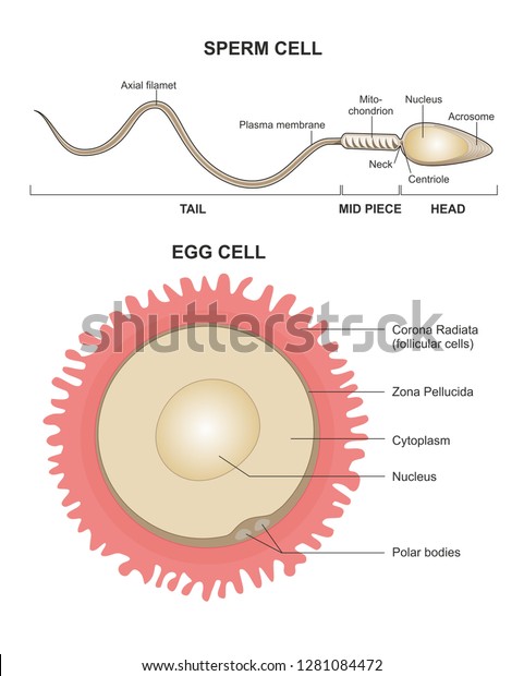
Spermatozoa are the only human cells that contain flagella. They are made up of three basic parts : the head, the middle-piece, and the tail.
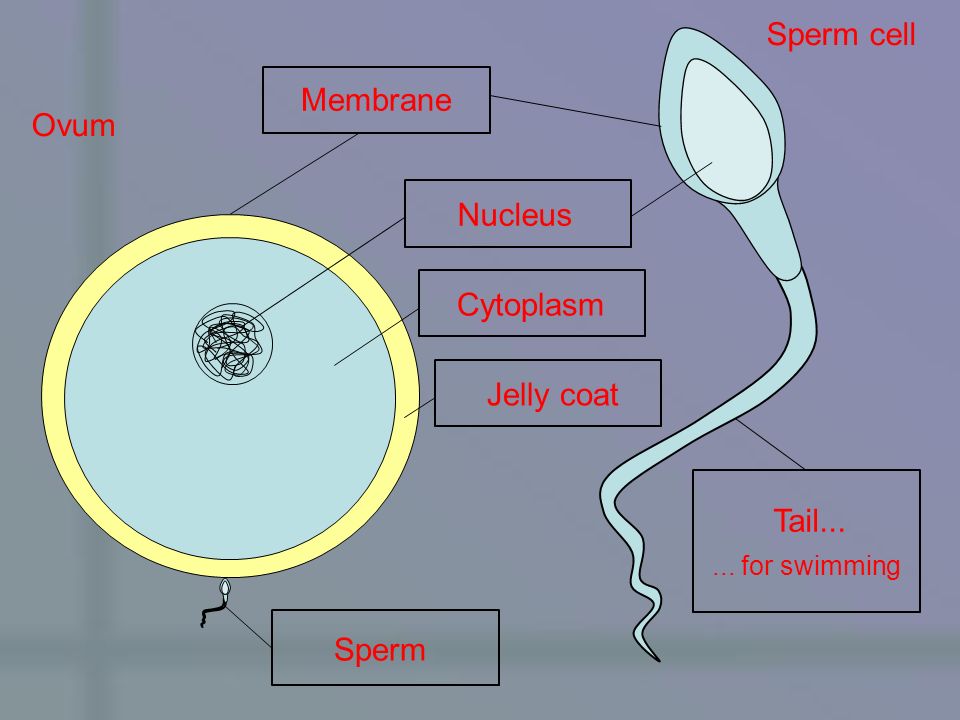
The head is an oval-shaped structure, which size ranges from 5 to 8 µm. It consists of two parts:
The neck and the middle piece, as the name suggests, are the parts that can be found between the head and the tail. Their function is to connect both ends of the sperm cell.
As one shall she in the diagram below, the neck contains millions of spirally arranged mitochondria . Their function is to provide the sperm will all the energy required by the flagellum to allow it to swim in the female reproductive tract.
The tail, also known as flagellum , is a long structure which main function is to allow sperm motility by means of a slithering, snake-like movement.
The length of the tail is about 50 µm, allowing a swimming velocity of 3 millimeters per minute approximately.
Sperm tail defects or alterations can lead to male fertility problems, being asthenozoospermia the most frequent one.
After ejaculation, most sperm die within minutes outside the woman's genital tract. If spermatozoa enter the female reproductive system (the cervix and uterus), they can survive 1-2 days , some up to 5 days.
Even though some sperms are able to survive for up to 5 days, almost all pregnancies can be attributed to intercourse that occurred 1-2 days prior to ovulation , as the percentage of sperms that are able to survive less than five days is higher.
On the other hand, when sperm are processed and stored under strict laboratory conditions in a nutrient-rich medium, they can remain alive for up to 7 days. If collected at home into a sterile container, their fertilizing capability will drop dramatically within 60 minutes.
So, in conclusion, the lifespan of human spermatozoa is 24-48 hours, but it depends on the environment, which is to say, the conditions under which they are held. If exposed to air or deposited on clothing, they dry out rapidly and die within minutes after ejaculation.
Sperm cell diseases or defects are more common than one may think. They occur in the structure of the sperm cell and can lead to male infertility, thereby preventing fertilization from taking place.
Teratozoospermia is a common abnormality that affects sperm morphology , that is, the shape of the sperm cells is altered (for example, sperm cells with 2 heads). Defects can be found either in the head, neck, tail, or in several parts simultaneously.
The sperm cell uses flagella to move towards the Fallopian tube; if they are damaged or not working, it will never meet the egg and subsequently fertilization will not occur. When male fertility issues are caused by alterations related to sperm motility, the patient is diagnosed with asthenozoospermia through a sperm analysis (SA). Vitamin supplements are often recommended as a home remedy when a man has issues related with the motility of his sperm.
Sperm diseases related to the anatomy of spermatozoa can translate into a man being infertile in the most severe cases. They can be caused by defects during spermatogenesis (sperm production), or by alterations acquired during the sperm maturation process and pathway until the sperms come out with ejaculation.
If you need to undergo IVF to become a mother, we recommend that you generate your Fertility Report now. In 3 simple steps, it will show you a list of clinics that fit your preferences and meet our strict quality criteria. Moreover, you will receive a report via email with useful tips to visit a fertility clinic for the first time.
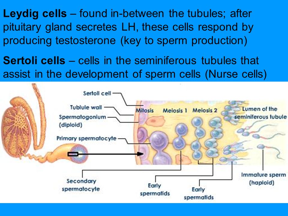
Spermatogenesis is the process whereby male reproductive cells are formed, from the immature ones, spermatogonia, until the mature ones, spermatozoa. This complicated process occurs within the seminiferous tubule in the testis and takes about 64-72 days.
Once spermatozoa (sperm cells) have been produced, they leave the testis and travel to the epididymis, where they will acquire the necessary motility in a process that lasts 10 days approximately. Spermatozoa will be stored in the epididymis until they are expelled with ejaculation. When ejaculation starts, sperm travel through the vasa deferentia and mingle with the seminal fluid that originates in the secretory glands, creating what we all know as semen . Finally, it is expelled through the urethra.
Sperm cells cannot divide because they are haploid, that is, they contain 23 chromosomes. However, after the fusion with the egg cell, which contains 23 chromosomes as well, they create a diploid cell of 46 chromosomes (totipotent zygote).
A spermatozoon (in plural spermatozoa ) is a motile sperm cell, that is, the reproductive cell of human males, carried in semen, that fertilizes the ovum to create a new human being, while the term sperm is commonly used to refer to the semen.
Approximately, the distance that spermatozoa travel through the female reproductive tract is about 15 to 18 cm. Experts have discovered that sperms have to travel distances that are around 1,000 times their own length while they swim in the right direction towards the egg.
Mitochondria supply energy to the sperm cells and are responsible for carrying out the process of respiration, necessary to provide the tail (flagellum) with the energy supply that it needed to allow the sperm cell to swim towards the egg.
As explained above, spermatozoa are haploid: they contain 23 chromosomes. Both sperm cells and egg cells contain half the chromosomes contained in normal diploid cells (a.k.a. somatic cells ). Haploid cells are produced during meiosis.
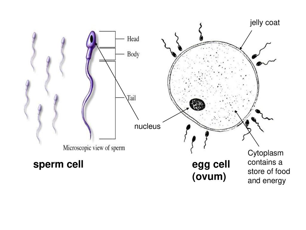
Egg cells are the complete opposite of sperm cells, actually. The main and most obvious difference is that egg cells are produced in the female reproductive system, while sperm cells develop in the testes, which are part of the male reproductive system.
On the other hands, while spermatozoa are one of the smallest cells in the male body, ova are one of the largest cells present in the body of human females.
Not literally, but in a way yes. Actually, blood brings oxygen and nutrition to the testicles. In fact, everything in our body (both in males and females) gets oxygen and nutrients from blood. So, while blood should not present in the semen, it is responsible for the sperm production process.
While there exist many old-wives tales and myths about this, the truth is that there is a 50% chance that a sperm cell will carry an X chromosome and a 50% chance that it will carry a Y chromosome. So, in short, the assignment of gender occurs 100% randomly.
We have laearned about the anatomy of sperm cells, but do you know how they are formed and where? This process, called spermatogenesis, is explained in detail here: How are sperm produced?
Now that we have learned about the parts and functions of a sperm cell, you may want to find out what is the pathway of sperm in their journey to the egg. Check this out to get more information: Sperm's journey to the egg .
Our editors have made great efforts to create this content for you. By sharing this post, you are helping us to keep ourselves motivated to work even harder.
Find the latest news on assisted reproduction in our channels.
Sperm Cells - Definition, Function, Structure, Adaptations, Microscopy
What’s the Function of a Sperm Cell ? - Definition & Structure
Sperm — Wikipedia Republished // WIKI 2 | Non-motile sperm cells
What Are Sperm Cells ? (with pictures)
Sperm cells (magnified 1,000 times). | Britannica
For other uses, see Sperm (disambiguation) .
Video of human sperm cells under a microscope
Sperm and egg fusing ( fertilisation )
Dimensions of the human sperm head measured from a 39 year-old healthy human subject.
Human sperm stained for semen quality testing
Further information: Sperm donation
Motile sperm cells of algae and seedless plants [31]
^ "Spermatium definition and meaning | Collins English Dictionary" . www.collinsdictionary.com . Retrieved 2020-02-20 .
^ Kumar, Anil (2006). Botany for Degree Gymnosperm (Multicolor ed.). S. Chand Publishing. p. 261. ISBN 978-81-219-2618-8 .
^ "Animal reproductive system - Male systems" . Encyclopedia Britannica . Retrieved 2020-02-20 .
^ Boitrelle, F; Guthauser, B; Alter, L; Bailly, M; Wainer, R; Vialard, F; Albert, M; Selva, J (2013). "The nature of human sperm head vacuoles: a systematic literature review" . Basic Clin Androl . 23 : 3. doi : 10.1186/2051-4190-23-3 . PMC 4346294 . PMID 25780567 .
^ Fawcett, D. W. (1981) Sperm Flagellum. In: The Cell. D. W. Fawcett. Philadelphia, W. B. Saunders Company. 14: pp. 604-640.
^ Lehti, M. S. and A. Sironen (2017). "Formation and function of sperm tail structures in association with sperm motility defects." Bi
^ Ishijima, Sumio; Oshio, Shigeru; Mohri, Hideo (1986). " Flagellar movement of human spermatozoa ". Gamete Research . 13 (3): 185–197. doi : 10.1002/mrd.1120130302 .
^ Wilson, Clare (July 31, 2020). "Sperm have a weird way of swimming and we only noticed after 300 years" . New Scientist .
^ Gadêlha, Hermes; Hernández-Herrera, Paul; Montoya, Fernando; Darszon, Alberto; Corkidi, Gabriel (July 2020). "Human sperm uses asymmetric and anisotropic flagellar controls to regulate swimming symmetry and cell steering" . Science Advances . 6 (31): eaba5168. doi : 10.1126/sciadv.aba5168 . PMC 7399739 . PMID 32789171 .
^ a b Fishman, Emily L; Jo, Kyoung; Nguyen, Quynh P. H; Kong, Dong; Royfman, Rachel; Cekic, Anthony R; Khanal, Sushil; Miller, Ann L; Simerly, Calvin; Schatten, Gerald; Loncarek, Jadranka; Mennella, Vito; Avidor-Reiss, Tomer (2018). "A novel atypical sperm centriole is functional during human fertilization" . Nature Communications . 9 (1): 2210. Bibcode : 2018NatCo...9.2210F . doi : 10.1038/s41467-018-04678-8 . PMC 5992222 . PMID 29880810 .
^ Blachon, S; Cai, X; Roberts, K. A; Yang, K; Polyanovsky, A; Church, A; Avidor-Reiss, T (2009). "A Proximal Centriole-Like Structure is Present in Drosophila Spermatids and Can Serve as a Model to Study Centriole Duplication" . Genetics . 182 (1): 133–44. doi : 10.1534/genetics.109.101709 . PMC 2674812 . PMID 19293139 .
^ Hewitson, Laura & Schatten, Gerald P. (2003). "The biology of fertilization in humans" . In Patrizio, Pasquale; et al. (eds.). A color atlas for human assisted reproduction: laboratory and clinical insights . Lippincott Williams & Wilkins. p. 3. ISBN 978-0-7817-3769-2 . Retrieved 2013-11-09 .
^ Semen and sperm quality
^ Gould, JE; Overstreet, JW; Hanson, FW (1984). "Assessment of human sperm function after recovery from the female reproductive tract" . Biology of Reproduction . 31 (5): 888–894. doi : 10.1095/biolreprod31.5.888 . PMID 6518230 .
^ Cyranoski, David (2016). "Researchers claim to have made artificial mouse sperm in a dish". Nature . doi : 10.1038/nature.2016.19453 . S2CID 87014225 .
^ Gurevich, Rachel (2008-06-10). "Does Age Affect Male Fertility?" . About.com . Retrieved 14 February 2010 .
^ Gavriliouk D, Aitken RJ (2015). "Damage to Sperm DNA Mediated by Reactive Oxygen Species: Its Impact on Human Reproduction and the Health Trajectory of Offspring". The Male Role in Pregnancy Loss and Embryo Implantation Failure . Advances in Experimental Medicine and Biology. 868 . pp. 23–47. doi : 10.1007/978-3-319-18881-2_2 . ISBN 978-3-319-18880-5 . PMID 26178844 .
^ Marchetti F, Wyrobek AJ (2008). "DNA repair decline during mouse spermiogenesis results in the accumulation of heritable DNA damage" . DNA Repair . 7 (4): 572–81. doi : 10.1016/j.dnarep.2007.12.011 . PMID 18282746 .
^ Marchetti F, Essers J, Kanaar R, Wyrobek AJ (2007). "Disruption of maternal DNA repair increases sperm-derived chromosomal aberrations" . Proceedings of the National Academy of Sciences of the United States of America . 104 (45): 17725–9. Bibcode : 2007PNAS..10417725M . doi : 10.1073/pnas.0705257104 . PMC 2077046 . PMID 17978187 .
^ Marchetti F, Bishop J, Gingerich J, Wyrobek AJ (2015). "Meiotic interstrand DNA damage escapes paternal repair and causes chromosomal aberrations in the zygote by maternal misrepair" . Scientific Reports . 5 : 7689. Bibcode : 2015NatSR...5E7689M . doi : 10.1038/srep07689 . PMC 4286742 . PMID 25567288 .
^ Lüpold, Stefan; Manier, Mollie K; Puniamoorthy, Nalini; Schoff, Christopher; Starmer, William T; Luepold, Shannon H. Buckley; Belote, John M; Pitnick, Scott (2016). "How sexual selection can drive the evolution of costly sperm ornamentation" . Nature . 533 (7604): 535–8. Bibcode : 2016Natur.533..535L . doi : 10.1038/nature18005 . PMID 27225128 . S2CID 4407752 .
^ Gardiner, Jennifer R (2016). "The bigger, the better" . Nature . 533 (7604): 476. doi : 10.1038/533476a . PMID 27225117 .
^ Sarfraz Manzoor (2 November 2012). "Come inside: the world's biggest sperm bank" . The Guardian . Retrieved 4 August 2013 .
^ a b c Assisted Reproduction in the Nordic Countries ncbio.org
^ a b FDA Rules Block Import of Prized Danish Sperm Posted Aug 13, 08 7:37 AM CDT in World, Science & Health
^ a b Steven Kotler (26 September 2007). "The God of Sperm" .
^ "Timeline: Assisted reproduction and birth control" . CBC News . Retrieved 2006-04-06 .
^ Fiedler, Anja; Rehdorf, Jessica; Hilbers, Florian; Johrdan, Lena; Stribl, Carola; Benecke, Mark (2008). "Detection of Semen (Human and Boar) and Saliva on Fabrics by a Very High Powered UV-/VIS-Light Source" . The Open Forensic Science Journal . 1 : 12–15. doi : 10.2174/1874402800801010012 .
^ Allery, J. P; Telmon, N; Mieusset, R; Blanc, A; Rougé, D (2001). "Cytological detection of spermatozoa: Comparison of three staining methods". Journal of Forensic Sciences . 46 (2): 349–51. doi : 10.1520/JFS14970J . PMID 11305439 .
^ Illinois State Police/President's DNA Initiative. "The Presidents's DNA Initiative: Semen Stain Identification: Kernechtrot" (PDF) . Retrieved 2009-12-10 .
^ a b c d e f Raven, Peter H.; Ray F. Evert; Susan E. Eichhorn (2005). Biology of Plants, 7th Edition . New York: W.H. Freeman and Company Publishers. ISBN 0-7167-1007-2 .
^ Bottino D, Mogilner A , Roberts T, Stewart M, Oster G (2002). "How nematode sperm crawl". Journal of Cell Science . 115 (Pt 2): 367–84. PMID 11839788 . CS1 maint: multiple names: authors list ( link )
^ Sumbali, Geeta (2005). The Fungi . Alpha Science Int'l Ltd. ISBN 1-84265-153-6 .
^ Maheshwari R (1999). "Microconidia of Neurospora crassa". Fungal Genetics and Biology . 26 (1): 1–18. doi : 10.1006/fgbi.1998.1103 . PMID 10072316 .
^ Avidor-Reiss, T; Khire, A; Fishman, EL; Jo, KH (2015). "Atypical centrioles during sexual reproduction" . Front Cell Dev Biol . 3 : 21. doi : 10.3389/fcell.2015.00021 . PMC 4381714 . PMID 25883936 .
^ Blachon, S.; Cai, X.; Roberts, K. A.; Yang, K.; Polyanovsky, A.; Church, A.; Avidor-Reiss, T. (May 2009). "A Proximal Centriole-Like Structure Is Present in Drosophila Spermatids and Can Serve as a Model to Study Centriole Duplication" . Genetics . 182 (1): 133–44. doi : 10.1534/genetics.109.101709 . PMC 2674812 . PMID 19293139 .
^ Avidor-Reiss, Tomer; Leroux, Michel R (2015). "Shared and Distinct Mechanisms of Compartmentalized and Cytosolic Ciliogenesis" . Current Biology . 25 (23): R1143–50. doi : 10.1016/j.cub.2015.11.001 . PMC 5857621 . PMID 26654377 .
This page was last edited on 29 November 2020, at 00:38
Basis of this page is in Wikipedia . Text is available under the CC BY-SA 3.0 Unported License . Non-text media are available under their specified licenses. Wikipedia® is a registered trademark of the Wikimedia Foundation, Inc. WIKI 2 is an independent company and has no affiliation with Wikimedia Foundation.
To install click the Add extension button. That's it.
The source code for the WIKI 2 extension is being checked by specialists of the Mozilla Foundation, Google, and Apple. You could also do it yourself at any point in time.
Sperm is the male reproductive cell , or gamete , in anisogamous forms of sexual reproduction (forms in which there is a larger, "female" reproductive cell and a smaller, "male" one). Animals produce motile sperm with a tail known as a flagellum , which are known as spermatozoa , while some red algae and fungi produce non-motile sperm cells, known as spermatia . [1] Flowering plants contain non-motile sperm inside pollen , while some more basal plants like ferns and some gymnosperms have motile sperm. [2]
Sperm cells form during the process known as spermatogenesis , which in amniotes ( reptiles and mammals ) takes place in the seminiferous tubules of the testes . [3] This process involves the production of several successive sperm cell precursors, starting with spermatogonia , which differentiate into spermatocytes . The spermatocytes then undergo meiosis , reducing their chromosome number by half, which produces spermatids . The spermatids then mature and, in animals, construct a tail, or flagellum, which gives rise to the mature, motile sperm cell. This whole process occurs constantly and takes around 3 months from start to finish.
Sperm cells cannot divide and have a limited lifespan, but after fusion with egg cells during fertilisation , a new organism begins developing, starting as a totipotent zygote . The human sperm cell is haploid , so that its 23 chromosomes can join the 23 chromosomes of the female egg to form a diploid cell. In mammals , sperm is stored in the epididymis and is released from the penis during ejaculation in a fluid known as semen .
The word sperm is derived from the Greek word σπέρμα , sperma , meaning "seed".
The main sperm function is to reach the ovum and fuse with it to deliver two sub-cellular structures: (i) the male pronucleus that contains the genetic material and (ii) the centrioles that are structures that help organize the microtubule cytoskeleton .
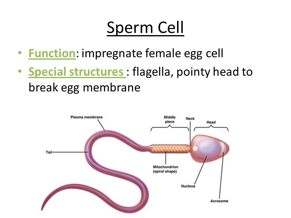
The mammalian sperm cell can be divided in 2 parts:
The neck or connecting piece contains one typical centriole and one atypical centriole such as the proximal centriole-like . [10] [11] The midpiece has a central filamentous core with many mitochondria spiralled around it, used for ATP production for the journey through the female cervix , uterus and uterine tubes .
During fertilization , the sperm provides three essential parts to the oocyte : (1) a signalling or activating factor, which causes the metabolically dormant oocyte to activate; (2) the haploid paternal genome ; (3) the centriole , which is responsible for forming the centrosome and microtubule system. [12]
The spermatozoa of animals are produced through spermatogenesis inside the male gonads ( testicles ) via meiotic division. The initial spermatozoon process takes around 70 days to complete. The process starts with the production of spermatogonia from germ cell precursors. These divide and differentiate into spermatocytes , which undergo meiosis to form spermatids . In the spermatid stage, the sperm develops the familiar tail. The next stage where it becomes fully mature takes around 60 days when it is called a spermatozoan . [13] Sperm cells are carried out of the male body in a fluid known as semen . Human sperm cells can survive within the female reproductive tract for more than 5 days post coitus. [14] Semen is produced in the seminal vesicles , prostate gland and urethral glands .
In 2016, scientists at Nanjing Medical University claimed they had produced cells resembling mouse spermatids from mouse embryonic stem cells artificially. They injected these spermatids into mouse eggs and produced pups. [15]
Sperm quantity and quality are the main parameters in semen quality , which is a measure of the ability of semen to accomplish fertilization . Thus, in humans, it is a measure of fertility in a man . The genetic quality of sperm, as well as its volume and motility, all typically decrease with age. [16] (See paternal age effect .)
DNA damages present in sperm cells in the period after meiosis but before fertilization may be repaired in the fertilized egg, but if not repaired, can have serious deleterious effects on fertility and the developing embryo. Human sperm cells are particularly vulnerable to free radical attack and the generation of oxidative DNA damage. [17] (see e.g. 8-Oxo-2'-deoxyguanosine )
The postmeiotic phase of mouse spermatogenesis is very sensitive to environmental genotoxic agents, because as male germ cells form mature sperm they progressively lose the ability to repair DNA damage. [18] Irradiation of male mice during late spermatogenesis can induce damage that persists for at least 7 days in the fertilizing sperm cells, and disruption of maternal DNA double-strand break repair pathways increases sperm cell-derived chromosomal aberrations. [19] Treatment of male mice with melphalan , a bifunctional alkylating agent frequently employed in chemotherapy, induces DNA lesions during meiosis that may persist in an unrepaired state as germ cells progress through DNA repair-competent phases of spermatogenic development. [20] Such unrepaired DNA damages in sperm cells, after fertilization, can lead to offspring with various abnormalities.
Related to sperm quality is sperm size, at least in some animals. For instance, the sperm of some species of fruit fly ( Drosophila ) are up to 5.8 cm long — about 20 times as long as the fly itself. Longer sperm cells are better than their shorter counterparts at displacing competitors from the female's seminal receptacle. The benefit to females is that only healthy males carry ‘good’ genes that can produce long sperm in sufficient quantities to outcompete their competitors. [21] [22]
Some sperm banks hold up to 170 litres (37 imp gal; 45 US gal) of sperm. [23]
In addition to ejaculation , it is possible to extract sperm through TESE .
On the global market, Denmark has a well-developed system of human sperm export. This success mainly comes from the reputation of Danish sperm donors for being of high quality [24] and, in contrast with the law in the other Nordic countries, gives donors the choice of being either anonymous or non-anonymous to the receiving couple. [24] Furthermore, Nordic sperm donors tend to be tall and highly educated [25] and have altruistic motives for their donations, [25] partly due to the relatively low monetary compensation in Nordic countries. More than 50 countries worldwide are importers of Danish sperm, including Paraguay , Canada , Kenya , and Hong Kong . [24] However, the Food and Drug Administration (FDA) of the US has banned import of any sperm, motivated by a risk of transmission of Creutzfeldt–Jakob disease , although such a risk is insignificant, since artificial insemination is very different from the route of transmission of Creutzfeldt–Jakob disease . [26] The prevalence of Creutzfeldt–Jakob disease for donors is at most one in a million, and if the donor was a carrier, the infectious proteins would still have to cross the blood-testis barrier to make transmission possible. [26]
Sperm were first observed in 1677 by Antonie van Leeuwenhoek [27] using a microscope . He described them as being animalcules (little animals), probably due to his belief in preformationism , which thought that each sperm contained a fully formed but small human. [ citation needed ]
Ejaculated fluids are detected by ultraviolet light , irrespective of the structure or colour of the surface. [28] Sperm heads, e.g. from vaginal swabs, are still detected by microscopy using the "Christmas Tree Stain" method, i.e., Kernechtrot-Picroindigocarmine (KPIC) staining. [29] [30]
Sperm cells in algal and many plant gametophytes are produced in male gametangia ( antheridia ) via mitotic division. In flowering plants , sperm nuclei are produced inside pollen . [ citation needed ]
Motile sperm cells typically move via flagella and require a water medium in order to swim toward the egg for fertilization. In animals most of the energy for sperm motility is derived from the metabolism of fructose carried in the seminal fluid . This takes place in the mitochondria located in the sperm's midpiece (at the base of the sperm head). These cells cannot swim backwards due to the nature of their propulsion. The uniflagellated sperm cells (with one flagellum) of animals are referred to as spermatozoa , and are known to vary in size. [ citation needed ]
Motile sperm are also produced by many protists and the gametophytes of bryophytes , ferns and some gymnosperms such as cycads and ginkgo . The sperm cells are the only flagellated cells in the life cycle of these plants. In many ferns and lycophytes , cycads and ginkgo they are multi-flagellated (carrying more than one flagellum). [31]
In nematodes , the sperm cells are amoeboid and crawl, rather than swim, towards the egg cell. [32]
Non-motile sperm cells called spermatia lack flagella and therefore cannot swim. Spermatia are produced in a spermatangium . [31]
Because spermatia cannot swim, they depend on their environment to carry them to the egg cell. Some red algae , such as Polysiphonia , produce non-motile spermatia that are spread by water currents after their release. [31] The spermatia of rust fungi are covered with a sticky substance. They are produced in flask-shaped structures containing nectar , which attract flies that transfer the spermatia to nearby hyphae for fertilization in a mechanism similar to insect pollination in flowering plants . [33]
Fungal spermatia (also called pycniospores, especially in the Uredinales) may be confused with conidia . Conidia are spores that germinate independently of fertilization, whereas spermatia are gametes that are required for fertilization. In some fungi, such as Neurospora crassa , spermatia are identical to microconidia as they can perform both functions of fertilization as well as giving rise to new organisms without fertilization. [34]
In almost all embryophytes , including most gymnosperms and all angiosperms , the male gametophytes ( pollen grains ) are the primary mode of dispersal , for example via wind or insect pollination , eliminating the need for water to bridge the gap between male and female. Each pollen grain contains a spermatogenous (generative) cell. Once the pollen lands on the stigma of a receptive flower, it germinates and starts growing a pollen tube through the carpel . Before the tube reaches the ovule , the nucleus of the generative cell in the pollen grain divides and gives rise to two sperm nuclei, which are then discharged through the tube into the ovule for fertilization. [31]
In some protists , fertilization also involves sperm nuclei , rather than cells, migrating toward the egg cell through a fertilization tube. Oomycetes form sperm nuclei in a syncytical antheridium surrounding the egg cells. The sperm nuclei reach the eggs through fertilization tubes, similar to the pollen tube mechanism in plants. [31]
Most sperm cells have centrioles in the sperm neck. [35] Sperm of many animals has 2 typical centrioles known as the proximal centriole and distal centriole. Some animals like human and bovine have a single typical centriole, known as the proximal centriole, and a second centriole with atypical structure. [10] Mice and rats have no recognizable sperm centrioles. The fruit fly Drosophila melanogaster has a single centriole and an atypical centriole named the Proximal Centriole-Like (PCL). [36]
The sperm tail is a specialized type of cilium (aka flagella). In many animals the sperm tail is formed in a unique way, which is named Cytosolic ciliogenesis , since all or part of axoneme of the sperm tail is formed in the cytoplasm or get exposed to the cytoplasm. [37]
Wikimedia Commons has media related to Sperm .
Rough Anal Sex Video
Censored Softcore Erotic Exoporn
Teen Peeing Video
Rise Private
Hd Girls Solo Orgasm


.jpg)