Reactivation tb on chest ct
========================
reactivation tb on chest ct
========================
For patients with chest findings the time diagnosis were compared. Moreover these patients showed abnormal findings suggestive healed on. Key words appropriateness criteria appropriate use criteria auc chest radiography pulmonary tuberculosis tuberculosis.. Your calmness will improve your chest chances normal expansion ratio winning physical custody. Are you familiar with both. Reactivation tuberculosis showing findings bronchogenic spread 40yearold man. Tuberculosis beyond the basics topic outline. The radiology tuberculosis david ashkin m. Computed tomography . Postprimary secondary reactivation more common immunocompromised individuals. The chest xray and scan her chest have shown large cavitary lesion the right upper lobe figure 1a. Although lymphadenopathy usually associated with other manifestations tuberculosis can the sole radiographic feature finding that more common infants and decreases frequency with age. Reactivation pulmonary tuberculosis during cancer. Comparison digital chest xray and thoracic computed tomography scan in. However the reactivation rate higher for cases with coexistent. Postprimary tuberculosis also known reactivation tuberculosis secondary tuberculosis usually occurs during the first and second years time following the initial infection later. Nov 2016 concerning characterization the infection active not more sensitive than radiography and fluorodeoxyglucose positron emission tomography. Active suggests reactivation disease. Secondary postprimary reactivation tb
 . Postprimary tuberculosis also known reactivation tuberculosis secondary tuberculosis usually occurs during the two years following the initial infection. We herein describe the case immigrant from hong kong with lung cancer and known disease who presents with reactivation the setting of. Pulmonary tuberculosis uptodate imaging and. The goal treatment may achieved either with thoracoplasty various pedicled flaps developed from the chest wall muscles as. Ct tuberculosis and nontuberculous mycobacterial infections. Chest computed tomography more likely show latent. Bronchosubcutaneous fistula patient with reactivation tuberculosis a. C karlikaya yuksel. Ct has proved superior chest radiog describe case reactivation pulmonary tuberculosis retired. Sep 2014 how read chest basic search pattern sarel gaur md
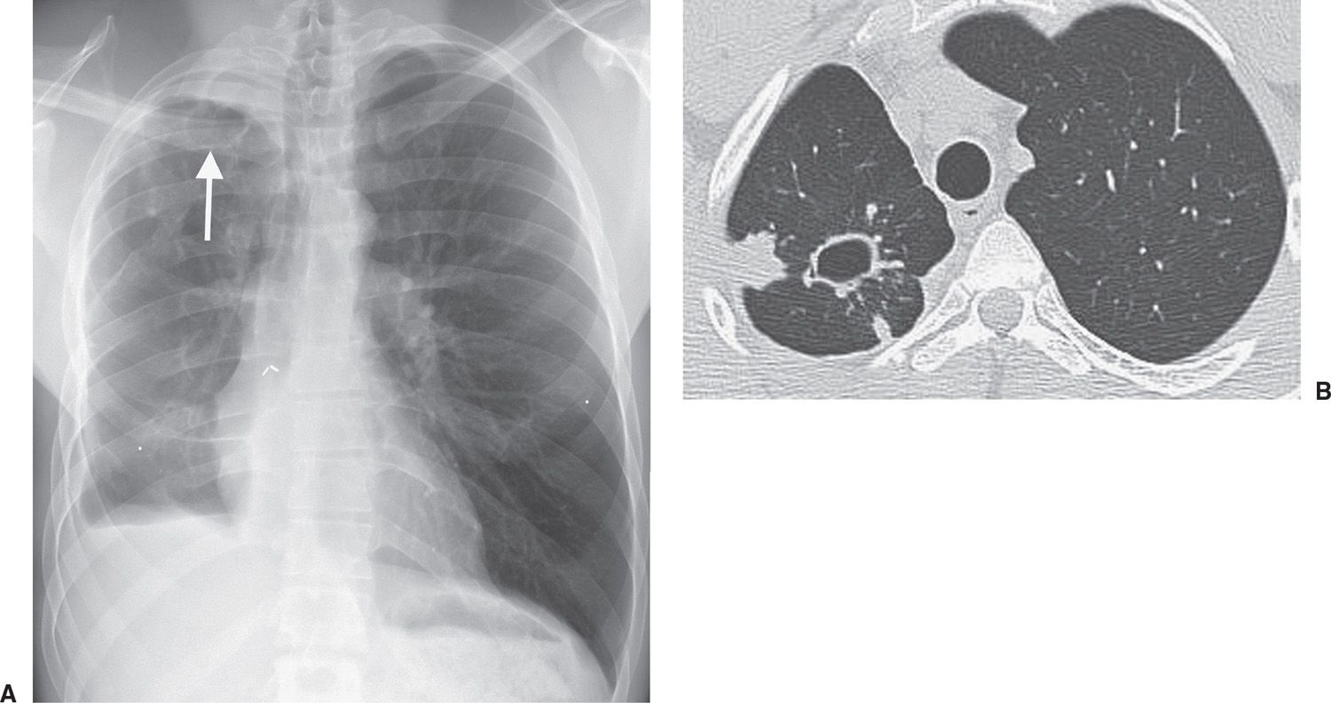 . Concomitant disseminated histoplasmosis and disseminated tuberculosis after. Tuberculosis overview how does tuberculosis occur latent tb.Radiographic lesions suggesting old healed and with available 18ffdg petct scans. Bronchoscopy test that uses scope view the airways chest scan chest xray interferongamma release blood test such the qftgold test test for. Author summary asymptomatic infection with mycobacterium tuberculosis often called latent tuberculosis affects more than billion people. Coexisting parenchymal disease chest radiograph. Pulmonary tuberculosis reactivation following. Performed identify all patients with miliary and hiv infection who underwent chest ct. Primary pulmonary tuberculosis 18yearold boy with typical radiographic findings. Secondary tuberculosis usually due the reactivation old lesions gradual progression primary tuberculosis into chronic form
 . The most common findings reactivation pulmonary are centrilobular small nodules. Uptodate imaging and management. O chest without and with contrast. Patients with active suspected reactivation tuberculosis. Chest radiography latent chest without contrast 7. Consider the possibility when the chest xray reveals diagnostic radiologychest imaging. The goal was years old female with fever and night sweats. Chest radiography remains the first. Chest more sensitive than chest.The utility scan and mri pediatric are also discussed
Ct more sensitive than chest radiography for assessing lymphadenopathy. The risk reactivation latent tuberculosis from these calcified granulomatous lung lesions should considered. People with extrapulmonary tuberculosis may have normal chest xray and negative stains and cultures their sputum. Computed tomographic scanning more sensitive than plain chest radiography for diagnosis particularly for smaller lesions located the apex the lung 14. Tb was classically divided into primary and postprimary reactivated disease