Per Vagina

👉🏻👉🏻👉🏻 ALL INFORMATION CLICK HERE 👈🏻👈🏻👈🏻
This article is about the birth canal. For the external female sex organs, see vulva. For other uses, see Vagina (disambiguation).
In mammals, the vagina is the elastic, muscular part of the female genital tract. In humans, it extends from the vulva to the cervix. The outer vaginal opening is normally partly covered by a membrane called the hymen. At the deep end, the cervix (neck of the uterus) bulges into the vagina. The vagina allows for sexual intercourse and birth. It also channels menstrual flow (menses), which occurs in humans and closely related primates as part of the monthly menstrual cycle.
Diagram of the female human reproductive tract and ovaries
superior part to uterine artery, middle and inferior parts to vaginal artery
uterovaginal venous plexus, vaginal vein
Sympathetic: lumbar splanchnic plexus
Parasympathetic: pelvic splanchnic plexus
Although research on the vagina is especially lacking for different animals, its location, structure and size are documented as varying among species. Female mammals usually have two external openings in the vulva, the urethral opening for the urinary tract and the vaginal opening for the genital tract. This is different from male mammals, who usually have a single urethral opening for both urination and reproduction. The vaginal opening is much larger than the nearby urethral opening, and both are protected by the labia in humans. In amphibians, birds, reptiles and monotremes, the cloaca is the single external opening for the gastrointestinal tract, the urinary, and reproductive tracts.
To accommodate smoother penetration of the vagina during sexual intercourse or other sexual activity, vaginal moisture increases during sexual arousal in human females and other female mammals. This increase in moisture provides vaginal lubrication, which reduces friction. The texture of the vaginal walls creates friction for the penis during sexual intercourse and stimulates it toward ejaculation, enabling fertilization. Along with pleasure and bonding, women's sexual behavior with others (which can include heterosexual or lesbian sexual activity) can result in sexually transmitted infections (STIs), the risk of which can be reduced by recommended safe sex practices. Other health issues may also affect the human vagina.
The vagina and vulva have evoked strong reactions in societies throughout history, including negative perceptions and language, cultural taboos, and their use as symbols for female sexuality, spirituality, or regeneration of life. In common speech, the word vagina is often used to refer to the vulva or to the female genitals in general. By its dictionary and anatomical definitions, however, vagina refers exclusively to the specific internal structure, and understanding the distinction can improve knowledge of the female genitalia and aid in healthcare communication.
The term vagina is from Latin meaning "sheath" or "scabbard"; the plural of vagina is either vaginae, or vaginas.[1] The vagina may also be referred to as "the birth canal" in the context of pregnancy and childbirth.[2][3] Although by its dictionary and anatomical definitions, the term vagina refers exclusively to the specific internal structure, it is colloquially used to refer to the vulva or to both the vagina and vulva.[4][5]
Using the term vagina to mean "vulva" can pose medical or legal confusion; for example, a person's interpretation of its location might not match another's interpretation of the location.[4][6] Medically, the vagina is the canal between the hymen (or remnants of the hymen) and the cervix, while, legally, it begins at the vulva (between the labia).[4] It may be that the incorrect use of the term vagina is due to not as much thought going into the anatomy of the female genitals as has gone into the study of male genitals, and that this has contributed to an absence of correct vocabulary for the external female genitalia among both the general public and health professionals. Because of this and because a better understanding of female genitalia can help combat sexual and psychological harm with regard to female development, researchers endorse correct terminology for the vulva.[6][7][8]
The human vagina is an elastic, muscular canal that extends from the vulva to the cervix.[9][10] The opening of the vagina lies in the urogenital triangle. The urogenital triangle is the front triangle of the perineum and also consists of the urethral opening and associated parts of the external genitalia.[11] The vaginal canal travels upwards and backwards, between the urethra at the front, and the rectum at the back. Near the upper vagina, the cervix protrudes into the vagina on its front surface at approximately a 90 degree angle.[12] The vaginal and urethral openings are protected by the labia.[13]
When not sexually aroused, the vagina is a collapsed tube, with the front and back walls placed together. The lateral walls, especially their middle area, are relatively more rigid. Because of this, the collapsed vagina has an H-shaped cross section.[10][14] Behind, the inner vagina is separated from the rectum by the recto-uterine pouch, the middle vagina by loose connective tissue, and the lower vagina by the perineal body.[15] Where the vaginal lumen surrounds the cervix of the uterus, it is divided into four continuous regions (vaginal fornices); these are the anterior, posterior, right lateral, and left lateral fornices.[9][10] The posterior fornix is deeper than the anterior fornix.[10]
Supporting the vagina are its upper, middle, and lower third muscles and ligaments. The upper third are the levator ani muscles, and the transcervical, pubocervical, and sacrocervical ligaments.[9][16] It is supported by the upper portions of the cardinal ligaments and the parametrium.[17] The middle third of the vagina involves the urogenital diaphragm.[9] It is supported by the levator ani muscles and the lower portion of the cardinal ligaments.[17] The lower third is supported by the perineal body,[9][18] or the urogenital and pelvic diaphragms.[19] The lower third may also be described as being supported by the perineal body and the pubovaginal part of the levator ani muscle.[16]
The vaginal opening is at the posterior end of the vulval vestibule, behind the urethral opening. The opening to the vagina is normally obscured by the labia minora (vaginal lips), but may be exposed after vaginal delivery.[10]
The hymen is a membrane of tissue that surrounds or partially covers the vaginal opening.[10] The effects of intercourse and childbirth on the hymen are variable. Where it is broken, it may completely disappear or remnants known as carunculae myrtiformes may persist. Otherwise, being very elastic, it may return to its normal position.[20] Additionally, the hymen may be lacerated by disease, injury, medical examination, masturbation or physical exercise. For these reasons, virginity cannot be definitively determined by examining the hymen.[20][21]
The length of the vagina varies among women of child-bearing age. Because of the presence of the cervix in the front wall of the vagina, there is a difference in length between the front wall, approximately 7.5 cm (2.5 to 3 in) long, and the back wall, approximately 9 cm (3.5 in) long.[10][22] During sexual arousal, the vagina expands both in length and width. If a woman stands upright, the vaginal canal points in an upward-backward direction and forms an angle of approximately 45 degrees with the uterus.[10][18] The vaginal opening and hymen also vary in size; in children, although the hymen commonly appears crescent-shaped, many shapes are possible.[10][23]
The vaginal plate is the precursor to the vagina.[24] During development, the vaginal plate begins to grow where the fused ends of the paramesonephric ducts (Müllerian ducts) enter the back wall of the urogenital sinus as the sinus tubercle. As the plate grows, it significantly separates the cervix and the urogenital sinus; eventually, the central cells of the plate break down to form the vaginal lumen.[24] This usually occurs by the twenty to twenty-fourth week of development. If the lumen does not form, or is incomplete, membranes known as vaginal septae can form across or around the tract, causing obstruction of the outflow tract later in life.[24]
There are conflicting views on the embryologic origin of the vagina. The majority view is Koff's 1933 description, which posits that the upper two-thirds of the vagina originate from the caudal part of the Müllerian duct, while the lower part of the vagina develops from the urogenital sinus.[27][28] Other views are Bulmer's 1957's description that the vaginal epithelium derives solely from the urogenital sinus epithelium,[29] and Witschi's 1970 research, which reexamined Koff's description and concluded that the sinovaginal bulbs are the same as the lower portions of the Wolffian ducts.[28][30] Witschi's view is supported by research by Acién et al., Bok and Drews.[28][30] Robboy et al. reviewed Koff and Bulmer's theories, and support Bulmer's description in light of their own research.[29] The debates stem from the complexity of the interrelated tissues and the absence of an animal model that matches human vaginal development.[29][31] Because of this, study of human vaginal development is ongoing and may help resolve the conflicting data.[28]
The vaginal wall from the lumen outwards consists firstly of a mucosa of stratified squamous epithelium that is not keratinized, with a lamina propria (a thin layer of connective tissue) underneath it. Secondly, there is a layer of smooth muscle with bundles of circular fibers internal to longitudinal fibers (those that run lengthwise). Lastly, is an outer layer of connective tissue called the adventitia. Some texts list four layers by counting the two sublayers of the mucosa (epithelium and lamina propria) separately.[32][33]
The smooth muscular layer within the vagina has a weak contractive force that can create some pressure in the lumen of the vagina; much stronger contractive force, such as during childbirth, comes from muscles in the pelvic floor that are attached to the adventitia around the vagina.[34]
The lamina propria is rich in blood vessels and lymphatic channels. The muscular layer is composed of smooth muscle fibers, with an outer layer of longitudinal muscle, an inner layer of circular muscle, and oblique muscle fibers between. The outer layer, the adventitia, is a thin dense layer of connective tissue and it blends with loose connective tissue containing blood vessels, lymphatic vessels and nerve fibers that are between pelvic organs.[12][33][22] The vaginal mucosa is absent of glands. It forms folds (transverse ridges or rugae), which are more prominent in the outer third of the vagina; their function is to provide the vagina with increased surface area for extension and stretching.[9][10]
The epithelium of the ectocervix (the portion the uterine cervix extending into the vagina) is an extension of, and shares a border with, the vaginal epithelium.[35] The vaginal epithelium is made up of layers of cells, including the basal cells, the parabasal cells, the superficial squamous flat cells, and the intermediate cells.[36] The basal layer of the epithelium is the most mitotically active and reproduces new cells.[37] The superficial cells shed continuously and basal cells replace them.[10][38][39] Estrogen induces the intermediate and superficial cells to fill with glycogen.[39][40] Cells from the lower basal layer transition from active metabolic activity to death (apoptosis). In these mid-layers of the epithelia, the cells begin to lose their mitochondria and other organelles.[37][41] The cells retain a usually high level of glycogen compared to other epithelial tissue in the body.[37]
Under the influence of maternal estrogen, the vagina of a newborn is lined by thick stratified squamous epithelium (or mucosa) for two to four weeks after birth. Between then to puberty, the epithelium remains thin with only a few layers of cuboidal cells without glycogen.[39][42] The epithelium also has few rugae and is red in color before puberty.[4] When puberty begins, the mucosa thickens and again becomes stratified squamous epithelium with glycogen containing cells, under the influence of the girl's rising estrogen levels.[39] Finally, the epithelium thins out from menopause onward and eventually ceases to contain glycogen, because of the lack of estrogen.[10][38][43]
Flattened squamous cells are more resistant to both abrasion and infection.[42] The permeability of the epithelium allows for an effective response from the immune system since antibodies and other immune components can easily reach the surface.[44] The vaginal epithelium differs from the similar tissue of the skin. The epidermis of the skin is relatively resistant to water because it contains high levels of lipids. The vaginal epithelium contains lower levels of lipids. This allows the passage of water and water-soluble substances through the tissue.[44]
Keratinization happens when the epithelium is exposed to the dry external atmosphere.[10] In abnormal circumstances, such as in pelvic organ prolapse, the mucosa may be exposed to air, becoming dry and keratinized.[45]
Blood is supplied to the vagina mainly via the vaginal artery, which emerges from a branch of the internal iliac artery or the uterine artery.[9][46] The vaginal arteries anastamose (are joined) along the side of the vagina with the cervical branch of the uterine artery; this forms the azygos artery,[46] which lies on the midline of the anterior and posterior vagina.[15] Other arteries which supply the vagina include the middle rectal artery and the internal pudendal artery,[10] all branches of the internal iliac artery.[15] Three groups of lymphatic vessels accompany these arteries; the upper group accompanies the vaginal branches of the uterine artery; a middle group accompanies the vaginal arteries; and the lower group, draining lymph from the area outside the hymen, drain to the inguinal lymph nodes.[15][47] Ninety-five percent of the lymphatic channels of the vagina are within 3 mm of the surface of the vagina.[48]
Two main veins drain blood from the vagina, one on the left and one on the right. These form a network of smaller veins, the vaginal venous plexus, on the sides of the vagina, connecting with similar venous plexuses of the uterus, bladder, and rectum. These ultimately drain into the internal iliac veins.[15]
The nerve supply of the upper vagina is provided by the sympathetic and parasympathetic areas of the pelvic plexus. The lower vagina is supplied by the pudendal nerve.[10][15]
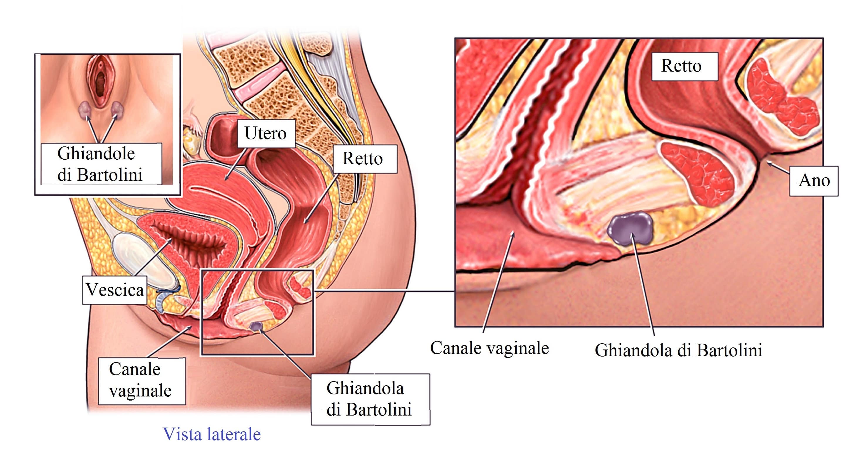
Vaginal secretions are primarily from the uterus, cervix, and vaginal epithelium in addition to minuscule vaginal lubrication from the Bartholin's glands upon sexual arousal.[10] It takes little vaginal secretion to make the vagina moist; secretions may increase during sexual arousal, the middle of or a little prior to menstruation, or during pregnancy.[10] Menstruation (also known as a "period" or "monthly") is the regular discharge of blood and mucosal tissue (known as menses) from the inner lining of the uterus through the vagina.[49] The vaginal mucous membrane varies in thickness and composition during the menstrual cycle,[50] which is the regular, natural change that occurs in the female reproductive system (specifically the uterus and ovaries) that makes pregnancy possible.[51][52] Different hygiene products such as tampons, menstrual cups, and sanitary napkins are available to absorb or capture menstrual blood.[53]
The Bartholin's glands, located near the vaginal opening, were originally considered the primary source for vaginal lubrication, but further examination showed that they provide only a few drops of mucus.[54] Vaginal lubrication is mostly provided by plasma seepage known as transudate from the vaginal walls. This initially forms as sweat-like droplets, and is caused by increased fluid pressure in the tissue of the vagina (vasocongestion), resulting in the release of plasma as transudate from the capillaries through the vaginal epithelium.[54][55][56]
Before and during ovulation, the mucus glands within the cervix secrete different variations of mucus, which provides an alkaline, fertile environment in the vaginal canal that is favorable to the survival of sperm.[57] Following menopause, vaginal lubrication naturally decreases.[58]
Nerve endings in the vagina can provide pleasurable sensations when the vagina is stimulated during sexual activity. Women may derive pleasure from one part of the vagina, or from a feeling of closeness and fullness during vaginal penetration.[59] Because the vagina is not rich in nerve endings, women often do not receive sufficient sexual stimulation, or orgasm, solely from vaginal penetration.[59][60][61] Although the literature commonly cites a greater concentration of nerve endings and therefore greater sensitivity near the vaginal entrance (the outer one-third or lower third),[60][61][62] some scientific examinations of vaginal wall innervation indicate no single area with a greater density of nerve endings.[63][64] Other research indicates that only some women have a greater density of nerve endings in the anterior vaginal wall.[63][65] Because of the fewer nerve endings in the vagina, childbirth pain is significantly more tolerable.[61][66][67]
Pleasure can be derived from the vagina in a variety of ways. In addition to penile penetration, pleasure can come from masturbation, fingering, oral sex (cunnilingus), or specific sex positions (such as the missionary position or the spoons sex position).[68] Heterosexual couples may engage in cunnilingus or fingering as forms of foreplay to incite sexual arousal or as accompanying acts,[69][70] or as a type of birth control, or to preserve virginity.[71][72] Less commonly, they may use non penile-vaginal sexual acts as a primary means of sexual pleasure.[70] By contrast, lesbians and other women who have sex with women commonly engage in cunnilingus or fingering as main forms of sexual activity.[73][74] Some women and couples use sex toys, such as a vibrator or dildo, for vaginal pleasure.[75] The Kama Sutra – an ancient Hindu text written by Vātsyāyana, which includes a number of sexual positions – may also be used to increase sexual pleasure,[76] with special emphasis on female sexual satisfaction.[77]
Most women require direct stimulation of the clitoris to orgasm.[60][61] The clitoris plays a part in vaginal stimulation. It is a sex organ of multiplanar structure containing an abundance of nerve endings, with a broad attachment to the pubic arch and extensive supporting tissue to the labia. Research indicates that it forms a tissue cluster with the vagina. This tissue is perhaps more extensive in some women than in others, which may contribute to orgasms experienced vaginally.[60][78][79
Cruel Handjob Porn
Jean Franco Xxx
He Fuck His Sister
Spanking Anal Porn
Petite Sterling
per vagina - Wiktionary
PV - Per Vagina - All Acronyms
Vagina - Wikipedia
The Perfect Vagina - video Dailymotion
B-Lady Vagina-Prothese, Vagina - Transgender Shop
Come Lavare la Tua Vagina: 15 Passaggi (con Immagini)
Erotische Fotos - FeedBurner
Come Mantenere il Buon Odore della Vagina: 15 Passaggi
Anna Swan: Die Frau mit der größten Vagina der Welt
Per Vagina