Pancreatic and Urinary disorders
Alex ZPancreas
Endocrine - secretion of digestive enzymes
Endocrine - consists of islets of Langerhans and secretes insulin, glucagon and gastrin
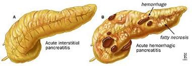
Acute Pancreatitis - release of pancreatic enzymes leading to necrosis
Etiology: Gall stones, trauma, alcohol and drugs
Symptoms: Discomfort, nausea, vomiting, pain
Sequelae: Resolution, pancreatic pseudocysts, fibrosis.
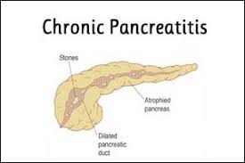
Chronic Pancreatitis - release of enzymes leading to necrosis
Aetiology: Alcohol abuse, gall stones, autoimmune disorders, cystic fibrosis
Symptoms: Persistant abdominal pain, steaterrhoe (due to malabsorbtion), jaundice
Carcinoma of the pancreas - typical adenocarcinoma
Aetiology: smoking, chronic pancreatitis, diabetes
Sites: 60% cases head of organ affected
Symptoms: late presentation and jaundace
Spread: Local spread, haematogenous (spread to liver)
Prognosis: Grave 5-year survival

Diagnosis of pancreatitis and pancreatic cancer
Acute pancreatitis
Amylase, lipase levels elevated
Chronic pancreatitis
MRI, endoscopic ultrasound
Fat in stool
Pancreatic cancer
CT, MRI, endoscopic ultrasound, PET scan, biopsy via ERCP
Diabetes mellitus
Disease develops due to disordered metabolism or due to lack of insulin in the body, leading to high blood glucose levels over prolonged period

Fasting blood glucose > 7.8 mmol/L
2h post-prandial blood glucose > 11.1 mmol.L
Urine: glycosuria, ketonuria, acetone in urine
Types
Type 1 - loss of insulin producing beta cells of the islet of Langerhans in the pancreas
Aetiology: autoimmune mediated
Insulin dependent, juvenile
Prognosis: increased risk of severe sight loss, end-stage kidney disease, cardiovascular dg.
Treatment:
A - long acting insulin injection,
B - regular meals and carbohydrates control,
C - use of insulin pump
Type 2 - insulin resistance and reduced insulin secretion
Aetiology: Increased age, obesity, polygenic inheritance and life factors
Non-insulin dependent, adult onset
Prognosis: increased risk of heart disease and stroke, diet control is an important factor
Treatment:
Diet, drug therapy
Gestational - developed by women during pregnancy
Secondary - developed due to other disease (e.g. cystic fibrosis)
Kidney affected (diabetic nephropathy)
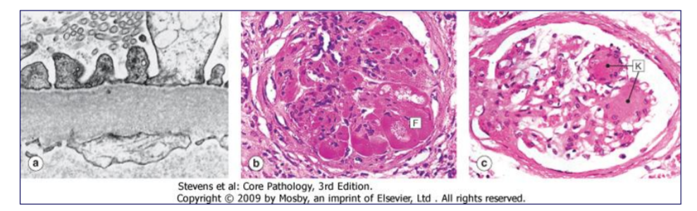
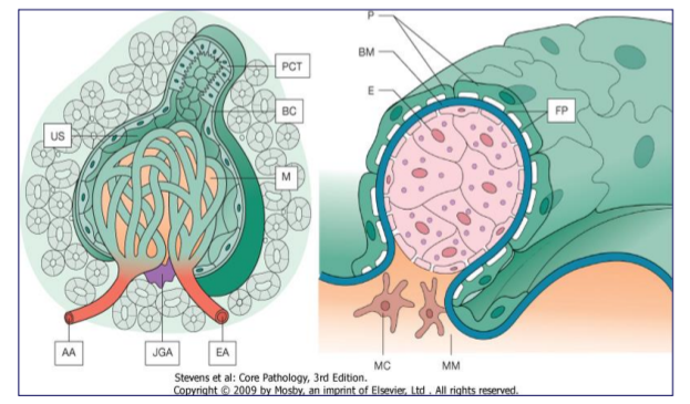
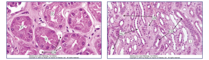
Renal failure - only some elements of renal function impared
Asymptomatic haematuria - early stage or minor glomerular damage
Persistent proteinuria - glomerular damage in early stages
Nephrotic syndrome - damage to glomerular basement membrane or mesangium, presented with heavy proteinuria, hypoalbuminemia, generalised oedema, hypercholesterolaemia, hyperlipidaemia
Nephritic syndrome - Abnormal glomerular structure with reactive cellular prolifiration, present with haematuria, oliguria, azotaemia, uraemia, oedema, mild hypertension
Total renal failure - all functions of nephron are impared
Acute - Central perfusion failure, (seen in hypovolemic shock & sudden profound hypotension), Presents with oliguria, anuria, disturbances in fluid & electrolyte balance
Chronic - Progressive destruction of nephrons over time by vascular, glomerular and/or tubular disease, congenital disease - (ADPKD)
~ Failure of renal activation of Vit D & increased loss of calcium in urine Destruction of renal parenchyma and anaemia
~ Detected by GFR (glomerular filtration rate)
Normal = 90-140mL/min
Chronic renal failure = 15-29 mL/min
End stage renal failure <14 mL/min
Glomerular disease
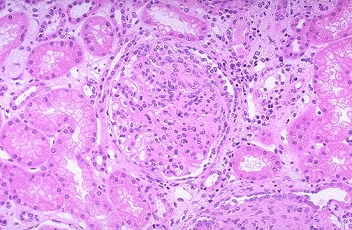
Deposition of immune complexes in the glomeruli, stimulated by preceding infection (Streptococcal spp. most common) Occurs in genetically predisposed patients 1-4 weeks after infection GN develops as a Type III hypersensitivity

Acute Pyelonephritis
Most common cause is bacterial infection with E. coli
Enter the kidney by two routes:
Ascending infection from the lower urinary tract - most common - due to pregnancy, diabetes mellitus, stasis of urine
Bloodstream spread in septicaemic states - unusual - mainly seen in elderly
Clinical features: Fever, chills, pain in the flank/back, lower UTI signs
Diagnosis: Examination of urine, esp. culture
Pathological features: Small, cortical abscesses (pus within tubules) Linear streaks in medulla (pus within collecting tubules & ducts)
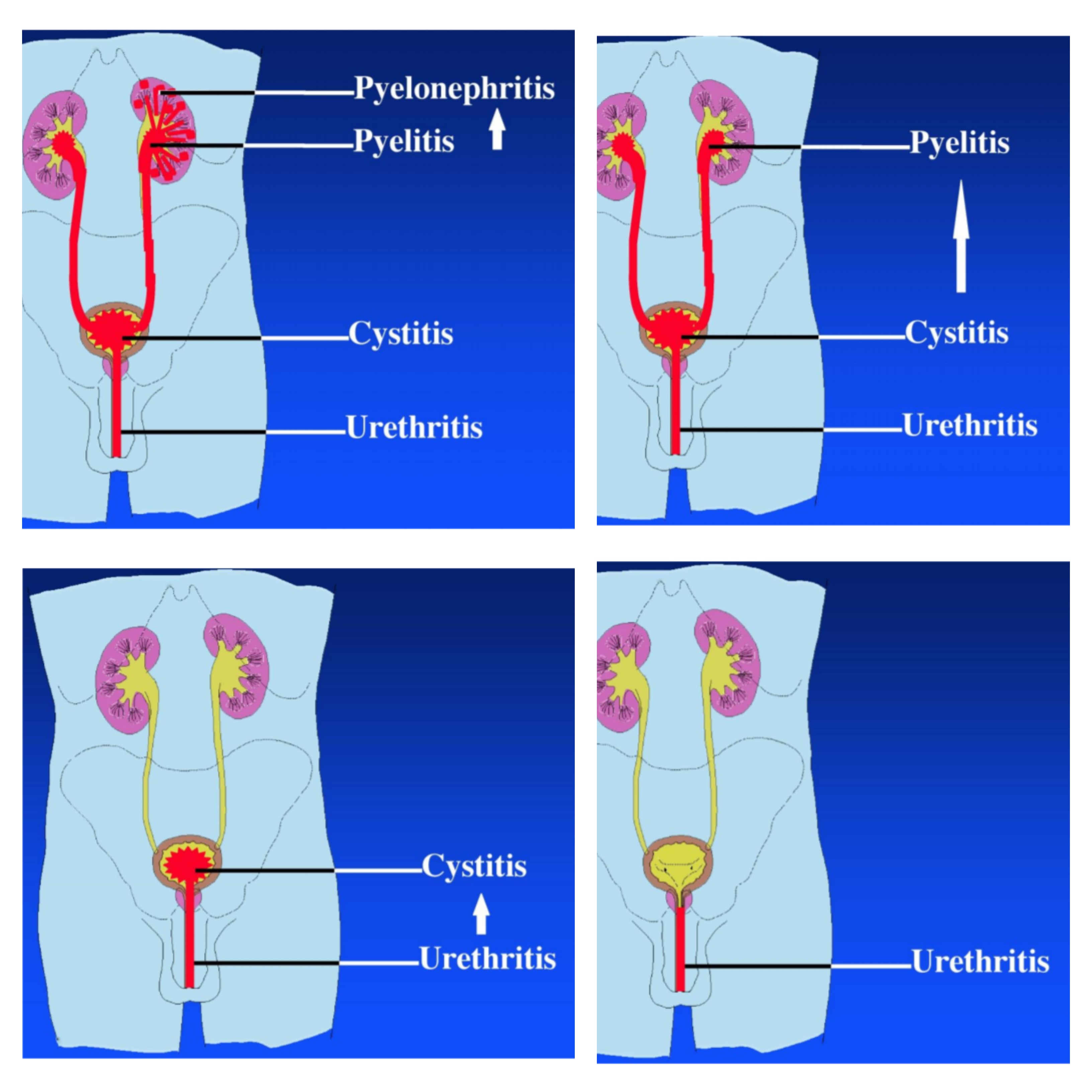
Chronic Pyelonephritis
Chronic inflammation of interstitium associated with irregular large scars of the kidney (nephrons destroyed) Most common site is renal calyces at poles of kidney Distortion of the pelvicalyceal system 2 o HT may develop vascular damage increases renal damage Common cause of end-stage chronic renal failure
Two Forms:
Reflux-Associated Chronic Pyelonephritis Reflux of urine from the bladder to ureters inflammation, fibrosis
Obstructive Chronic Pyelonephritis Obstruction of pelvicalyceal drainage (e.g. anatomical abnormality/stone) inflammation, fibrosis, recurrent renal infections
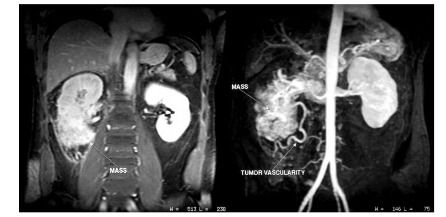
Renal Cell Carcinoma
Aetiology: Mainly idiopathic but some association with carcinogens (nitrosamines, cigarette smoking), chronic renal damage, genetic factors
Macroscopic features: Rounded masses with a yellowish cut face marked with areas of haemorrhage and necrosis
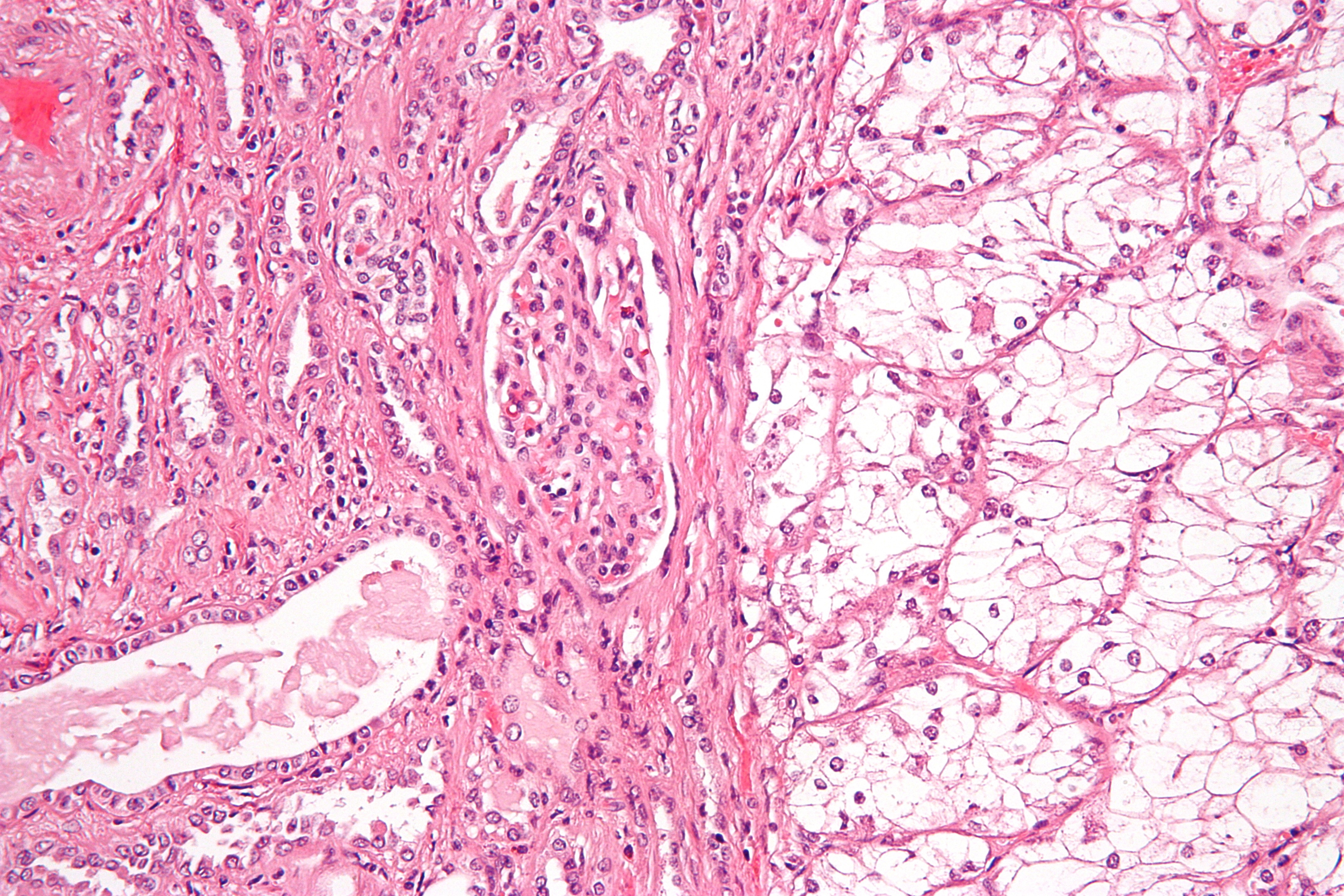
Histological patterns:
Clear cell carcinoma Most common > 70%
increased content of glycogen & lipid in cell cytoplasm
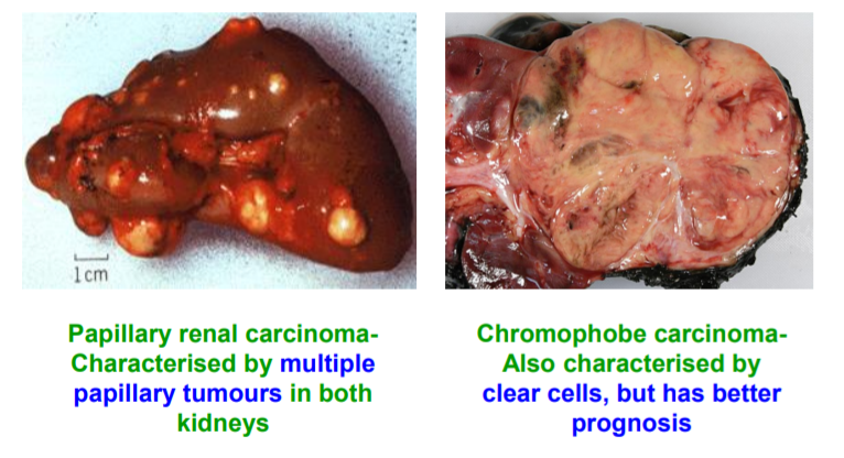
Papillary renal carcinoma (10-20%)
Chromophobe carcinoma (rarest)
Diagnosis by renal biopsy
Prognosis: 70% 5yr survival if confined within renal capsule Very poor if spread into perinephric fat, renal vein or metastases present (e.g. bone, brain, lung)
Summary
- Acute and chronic pancreatitis may have common aetiology, but the acute disease leads to much higher amylase, lipase levels
- Pancreatic adenocarcinoma has a very grave prognosis
- Type 1 and Type 2 DM have different aetiology and treatment methods, however diagnostic methods are similar, and symptoms and prognosis vary depending on effectiveness of treatment – both have multisystem manifestations
- Nephrotic (proteinuria) & nephritic (haematuria) syndromes are indicative of partial renal failure - glomerulonephritis may lead to both and can progress to acute or chronic renal failure
- Renal artery stenosis and chronic pyelonephritis may also lead to chronic renal failure
- Acute pyelonephritis commonly occurs due to an ascending infection from the lower urinary tract (E.coli) – pain in the flank/back – females more prone to UTIs
- Renal cell carcinoma presents with haematuria and loin pain and has a 70% 5 year survival rate if confined to the renal capsule
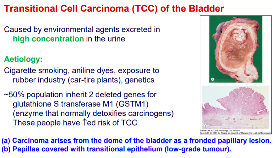
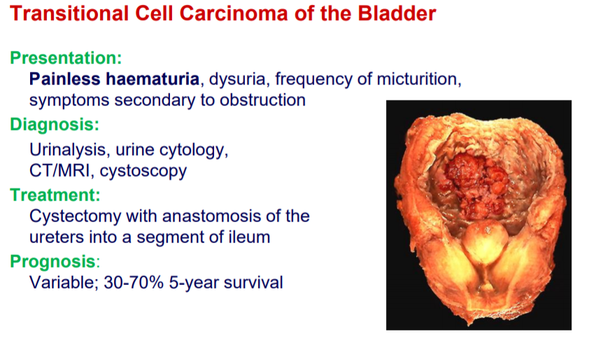
- TCC presents with painless haematuria and has a variable prognosis 30- 70% 5 year survival rate
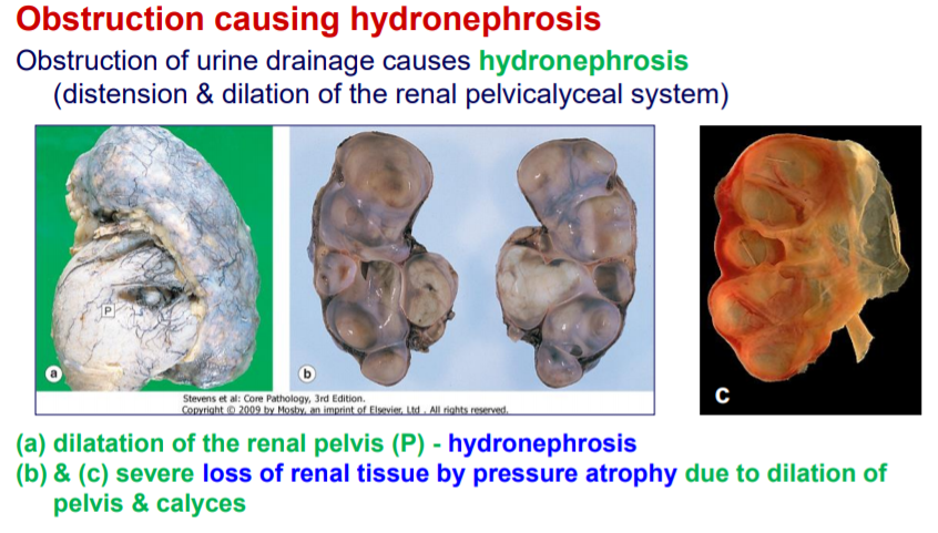
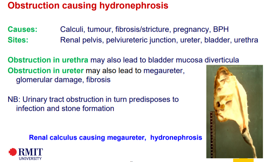
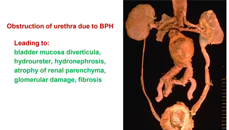
- Hydronephrosis is caused by factors that lead to obstruction of the lower urinary tract –which in turn cause infections and stone formation
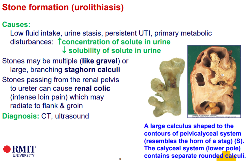
- Most common urinary stones are calcium oxalate and phosphate calculi – can cause renal colic
Extra stuff from slides