Mitral Valve Prolapse

💣 👉🏻👉🏻👉🏻 ALL INFORMATION CLICK HERE 👈🏻👈🏻👈🏻
Mitral valve prolapse and regurgitation
Associated Procedures
Cardiac catheterization
Chest X-rays
Coronary angiogram
Echocardiogram
Electrocardiogram (ECG or EKG)
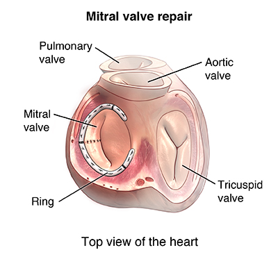
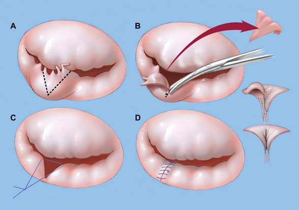
Heart valve surgery
Mitral valve repair and mitral valve replacement
Stress test
Show more associated procedures
© 1998-2021 Mayo Foundation for Medical Education and Research (MFMER). All rights reserved.
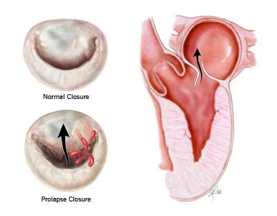
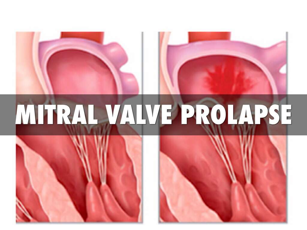
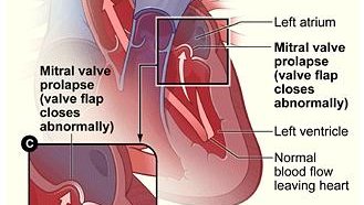
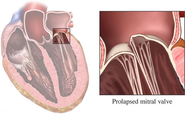
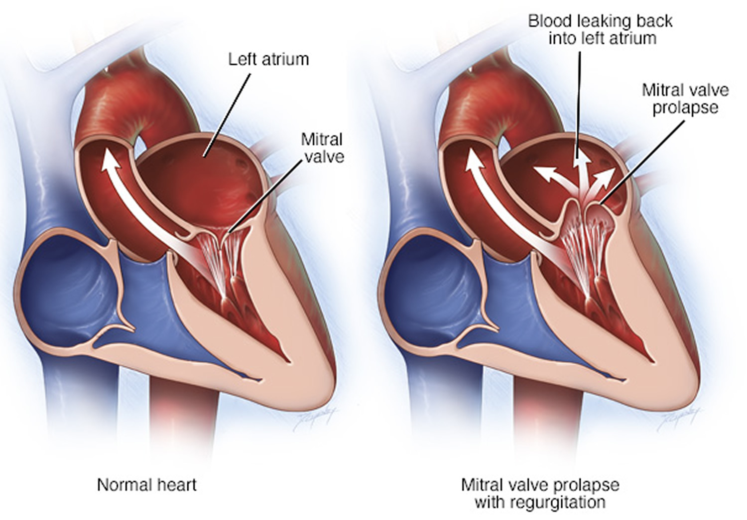
Mitral valve prolapse occurs when the flaps (leaflets) of the heart's mitral valve bulge (prolapse) like a parachute into the heart's left upper chamber (left atrium) as the heart contracts.
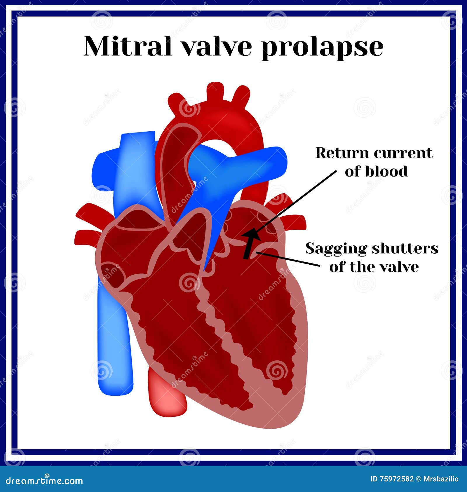
Mitral (MY-trul) valve prolapse sometimes leads to blood leaking backward into the left atrium, a condition called mitral valve regurgitation.
In most people, mitral valve prolapse isn't life-threatening and doesn't require treatment or changes in lifestyle. Some people with mitral valve prolapse, however, require treatment.
Although mitral valve prolapse is usually a lifelong disorder, many people with this condition never have symptoms. When diagnosed, people may be surprised to learn that they have a heart condition.
When signs and symptoms do occur, it may be because blood is leaking backward through the valve. Mitral valve prolapse symptoms can vary widely from one person to another. They tend to be mild and develop gradually. Symptoms may include:
If you think you have any of the above symptoms, make an appointment with your doctor. Many other conditions cause the same symptoms as mitral valve prolapse, so only a visit to your doctor can determine the cause of your symptoms.
If you're having chest pain and you're unsure if it could be a heart attack, seek emergency medical care immediately.
If you've already been diagnosed with mitral valve prolapse, see your doctor if your symptoms worsen.
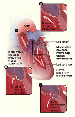
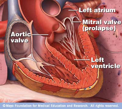
The mitral valve controls the flow of blood between the upper and lower chambers of the left side of the heart. When your heart is working properly, the mitral valve closes completely when the heart pumps and prevents blood from flowing back into the upper left chamber (left atrium).
But in some people with mitral valve prolapse, one or both of the mitral valve leaflets have extra tissue or stretch more than normal, which causes them to bulge like a parachute into the left atrium each time the heart contracts.
The bulging may keep the valve from closing tightly. In some cases, blood may leak backward through the valve (mitral valve regurgitation).
This may not cause problems if only a small amount of blood leaks back into the left atrium. More severe mitral valve regurgitation can cause symptoms such as shortness of breath, fatigue or lightheadedness.
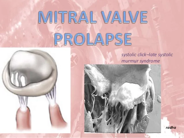
Another name for mitral valve prolapse is click-murmur syndrome. When a doctor listens to your heart using a stethoscope, he or she may hear a clicking sound as the valve's leaflets billow back, followed by a whooshing sound (murmur) resulting from blood flowing back into the atrium.
Other names to describe mitral valve prolapse include:
Mitral valve prolapse can develop in any person at any age. Serious symptoms of mitral valve prolapse tend to occur most often in men older than 50.
Mitral valve prolapse can run in families and may be linked to several other conditions, such as:
Although most people with mitral valve prolapse never have problems, complications can occur. They may include:
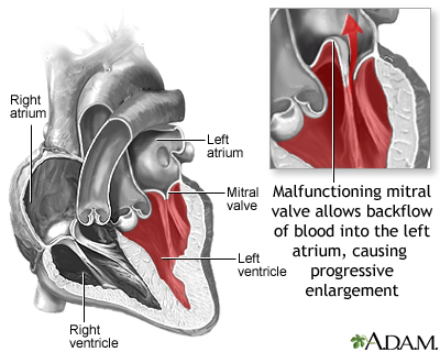
Mitral valve regurgitation. The most common complication is a condition in which the valve leaks blood back into the left atrium.
Being male or having high blood pressure increases your risk of mitral valve regurgitation.
If regurgitation is severe, you may need surgery to repair or replace the valve in order to prevent heart failure.
Heart rhythm problems (arrhythmias). Irregular heart rhythms most commonly occur in the upper chambers of the heart. They may be bothersome, but aren't usually life-threatening.
People with severe mitral valve regurgitation or severe deformity of their mitral valve are most at risk of having rhythm problems, which can affect blood flow through the heart.
Heart valve infection (endocarditis). The inside of your heart is lined by a thin membrane called the endocardium. Endocarditis is an infection of this inner lining.
An abnormal mitral valve increases your chance of getting endocarditis from bacteria, which can further damage the mitral valve.
Mayo Clinic does not endorse companies or products. Advertising revenue supports our not-for-profit mission.
Check out these best-sellers and special offers on books and newsletters from Mayo Clinic.
Any use of this site constitutes your agreement to the Terms and Conditions and Privacy Policy linked below.
A single copy of these materials may be reprinted for noncommercial personal use only. "Mayo," "Mayo Clinic," "MayoClinic.org," "Mayo Clinic Healthy Living," and the triple-shield Mayo Clinic logo are trademarks of Mayo Foundation for Medical Education and Research.
From Wikipedia, the free encyclopedia
Heart sounds of a 16-year-old girl diagnosed with mitral valve prolapse and mitral regurgitation. Auscultating her heart, a systolic murmur and click are heard. Recorded with the stethoscope over the mitral valve.
^ Jump up to: a b Barlow JB, Bosman CK (February 1966). "Aneurysmal protrusion of the posterior leaflet of the mitral valve. An auscultatory-electrocardiographic syndrome". Am. Heart J . 71 (2): 166–78. doi : 10.1016/0002-8703(66)90179-7 . PMID 4159172 .
^ Jump up to: a b c d e f Hayek E, Gring CN, Griffin BP (2005). "Mitral valve prolapse". Lancet . 365 (9458): 507–18. doi : 10.1016/S0140-6736(05)17869-6 . PMID 15705461 .
^ Criley JM, Lewis KB, Humphries JO, Ross RS (July 1966). "Prolapse of the mitral valve: clinical and cine-angiocardiographic findings" . Br Heart J . 28 (4): 488–96. doi : 10.1136/hrt.28.4.488 . PMC 459076 . PMID 5942469 .
^ Ahmed, Mustafa I.; Sanagala, Thriveni; Denney, Thomas; Inusah, Seidu; McGiffin, David; Knowlan, Donald; O’Rourke, Robert A.; Dell’Italia, Louis J. (August 2009). "Mitral Valve Prolapse With a Late-Systolic Regurgitant Murmur May Be Associated With Significant Hemodynamic Consequences". The American Journal of the Medical Sciences . 338 (2): 113–115. doi : 10.1097/MAJ.0b013e31819d5ec6 . PMID 19561453 . S2CID 44385990 .
^ Jump up to: a b Tanser, Paul H. (reviewed Mar 2007). "Mitral Valve Prolapse" , The Merck Manuals Online Medical Library , Retrieved 2011-01-08.
^ "Aortic Regurgitation" . The Lecturio Medical Concept Library . Retrieved 30 June 2021 .
^ Kolibash AJ (1988). "Progression of mitral regurgitation in patients with mitral valve prolapse". Herz . 13 (5): 309–17. PMID 3053383 .
^ "Related Disorders: Mitral Valve Prolapse" . Archived from the original on 2007-02-25 . Retrieved 2007-07-11 .
^ Lumiaho, A; Ikäheimo, R; Miettinen, R; Niemitukia, L; Laitinen, T; Rantala, A; Lampainen, E; Laakso, M; Hartikainen, J (2001). "Mitral valve prolapse and mitral regurgitation are common in patients with polycystic kidney disease type 1". American Journal of Kidney Diseases . 38 (6): 1208–16. doi : 10.1053/ajkd.2001.29216 . PMID 11728952 .
^ "Graves Disease" . www.niddk.nih.gov . August 10, 2012. Archived from the original on 30 June 2021 . Retrieved 2015-04-02 .
^ "Pectus Excavatum: Epidemiology" . Medscape . Retrieved 14 April 2016 .
^ Jump up to: a b c d e f g h Playford, David; Weyman, Arthur (2001). "Mitral valve prolapse: time for a fresh look" . Reviews in Cardiovascular Medicine . 2 (2): 73–81. PMID 12439384 . Archived from the original on 2014-09-03 . Retrieved 2009-03-24 .
^ Jump up to: a b Freed LA, Levy D, Levine RA, Larson MG, Evans JC, Fuller DL, Lehman B, Benjamin EJ (1999). "Prevalence and clinical outcome of mitral-valve prolapse". N Engl J Med . 341 (1): 1–7. doi : 10.1056/NEJM199907013410101 . PMID 10387935 .
^ Cotran, Ramzi S.; Kumar, Vinay; Fausto, Nelson; Nelso Fausto; Robbins, Stanley L.; Abbas, Abul K. (2005). Robbins and Cotran pathologic basis of disease . St. Louis, Mo: Elsevier Saunders. ISBN 978-0-7216-0187-8 . Archived from the original on 10 September 2005.
^ NLM/NIH: Medline Plus Medical Encyclopedia: Rheumatic fever
^ S Venkatesan; et al. (Sep–Oct 2007). "Can we diagnose Infective endocarditis without vegetation?" . Indian Heart Journal . 59 (5).
^ Gray's anatomy : the anatomical basis of clinical practice . Standring, Susan (Forty-first ed.). [Philadelphia]. 2016. ISBN 9780702052309 . OCLC 920806541 . CS1 maint: others ( link )
^ Toomer KA, Yu M, Fulmer D, Guo L, Moore KS, Moore R, Drayton KD, Glover J, Peterson N, Ramos-Ortiz S, Drohan A, Catching BJ, Stairley R, Wessels A, Lipschutz JH, Delling FN, Jeunemaitre X, Dina C, Collins RL1, Brand H10, Talkowski ME10, Del Monte F11, Mukherjee R, Awgulewitsch A, Body S, Hardiman G, Hazard ES, da Silveira WA, Wang B, Leyne M, Durst R, Markwald RR, Le Scouarnec S, Hagege A, Le Tourneau T, Kohl P, Rog-Zielinska EA, Ellinor PT, Levine RA, Milan DJ, Schott JJ, Bouatia-Naji N, Slaugenhaupt SA, Norris RA (2019) Primary cilia defects causing mitral valve prolapse. Sci Transl Med 11(493)
^ "Mitral valve prolapse" . Mayo Clinic.
^ "Mitral Regurgitation" . The Lecturio Medical Concept Library . Retrieved 11 August 2021 .
^ Wilson W, Taubert KA, Gewitz M, et al. (2007). "Prevention of infective endocarditis: guidelines from the American Heart Association" (PDF) . Journal of the American Dental Association . 138 (6): 739–45, 747–60. doi : 10.14219/jada.archive.2007.0262 . PMID 17545263 .
^ "Mitral Valve Prolapse" . The Lecturio Medical Concept Library . Retrieved 18 July 2021 .
^ Jump up to: a b Mitral Valve Prolapse at eMedicine
^ "Mitral Valve Prolapse" . The Lecturio Medical Concept Library . Retrieved 3 July 2021 .
^ Levy D, Savage D (1987). "Prevalence and clinical features of mitral valve prolapse". Am Heart J . 113 (5): 1281–90. doi : 10.1016/0002-8703(87)90956-2 . PMID 3554946 .
^ Warth DC, King ME, Cohen JM, Tesoriero VL, Marcus E, Weyman AE (May 1985). "Prevalence of mitral valve prolapse in normal children" . Journal of the American College of Cardiology . 5 (5): 1173–7. doi : 10.1016/S0735-1097(85)80021-8 . PMID 3989128 .
^ Barlow JB, Bosman CK (1966). "Aneurysmal protrusion of the posterior leaflet of the mitral valve. An auscultatory-electrocardiographic syndrome". Am Heart J . 71 (2): 166–78. doi : 10.1016/0002-8703(66)90179-7 . PMID 4159172 .
Wiki Loves Monuments: your chance to support Russian cultural heritage!
Photograph a monument and win!
Mitral valve prolapse ( MVP ) is a valvular heart disease characterized by the displacement of an abnormally thickened mitral valve leaflet into the left atrium during systole . [2] It is the primary form of myxomatous degeneration of the valve. There are various types of MVP, broadly classified as classic and nonclassic. In severe cases of classic MVP, complications include mitral regurgitation , infective endocarditis , congestive heart failure , and, in rare circumstances, cardiac arrest .
The diagnosis of MVP depends upon echocardiography , which uses ultrasound to visualize the mitral valve. MVP is estimated to affect 2–3% of the population. [2]
The condition was first described by John Brereton Barlow in 1966. [1] It was subsequently termed mitral valve prolapse by J. Michael Criley . [3]
Upon auscultation of an individual with mitral valve prolapse, a mid-systolic click, followed by a late systolic murmur heard best at the apex, is common. The length of the murmur signifies the time period over which blood is leaking back into the left atrium, known as regurgitation. A murmur that lasts throughout the whole of systole is known as a holo-systolic murmur. A murmur that is mid to late systolic, although typically associated with less regurgitation, can still be associated with significant hemodynamic consequences. [4]
In contrast to most other heart murmurs, the murmur of mitral valve prolapse is accentuated by standing and valsalva maneuver (earlier systolic click and longer murmur) and diminished with squatting (later systolic click and shorter murmur). The only other heart murmur that follows this pattern is the murmur of hypertrophic cardiomyopathy . An MVP murmur can be distinguished from a hypertrophic cardiomyopathy murmur by the presence of a mid-systolic click which is virtually diagnostic of MVP. The handgrip maneuver diminishes the murmur of an MVP and the murmur of hypertrophic cardiomyopathy. The handgrip maneuver also diminishes the duration of the murmur and delays the timing of the mid-systolic click. [5]
Both valsalva maneuver and standing decrease venous return to the heart thereby decreasing left ventricular diastolic filling ( preload ) and causing more laxity on the chordae tendineae . This allows the mitral valve to prolapse earlier in systole , leading to an earlier systolic click (i.e. closer to S 1 ), and a longer murmur. [6]
Historically, the term mitral valve prolapse syndrome has been applied to MVP associated with palpitations , atypical precordial pain , dyspnea on exertion , low body mass index , and electrocardiogram abnormalities ( ventricular tachycardia ), syncope , low blood pressure , headaches, lightheadedness , and other signs suggestive of autonomic nervous system dysfunction ( dysautonomia ). [2]
Mitral valve prolapse is frequently associated with mild mitral regurgitation , [7] where blood aberrantly flows from the left ventricle into the left atrium during systole . In the United States , MVP is the most common cause of severe, non-ischemic mitral regurgitation. [2] This is occasionally due to rupture of the chordae tendineae that support the mitral valve. [5]
MVP may occur with greater frequency in individuals with Ehlers-Danlos syndrome , Marfan syndrome [8] or polycystic kidney disease . [9] Other risk factors include Graves disease [10] and chest wall deformities such as pectus excavatum . [11] For unknown reasons, MVP patients tend to have a low body mass index (BMI) and are typically leaner than individuals without MVP. [12] [13]
Rheumatic fever is common worldwide and responsible for many cases of damaged heart valves . Chronic rheumatic heart disease is characterized by repeated inflammation with fibrinous resolution. The cardinal anatomic changes of the valve include leaflet thickening, commissural fusion, and shortening and thickening of the tendinous cords. [14] The recurrence of rheumatic fever is relatively common in the absence of maintenance of low dose antibiotics, especially during the first three to five years after the first episode. Heart complications may be long-term and severe, particularly if valves are involved. Rheumatic fever, since the advent of routine penicillin administration for Strep throat, has become less common in developed countries. In the older generation and in much of the less-developed world, valvular disease (including mitral valve prolapse, reinfection in the form of valvular endocarditis, and valve rupture) from undertreated rheumatic fever continues to be a problem. [15]
In an Indian hospital between 2004 and 2005, 4 of 24 endocarditis patients failed to demonstrate classic vegetations. All had rheumatic heart disease (RHD) and presented with prolonged fever. All had severe eccentric mitral regurgitation (MR). (One had severe aortic regurgitation (AR) also.) One had flail posterior mitral leaflet (PML). [16]
The mitral valve , so named because of its resemblance to a bishop 's mitre , is the heart valve that prevents the backflow of blood from the left ventricle into the left atrium of the heart. It is composed of two leaflets, one anterior and one posterior, that close when the left ventricle contracts. [17]
Each leaflet is composed of three layers of tissue : the atrialis , fibrosa , and spongiosa . Patients with classic mitral valve prolapse have excess connective tissue that thickens the spongiosa and separates collagen bundles in the fibrosa. This is due to an excess of dermatan sulfate , a glycosaminoglycan . This weakens the leaflets and adjacent tissue, resulting in increased leaflet area and elongation of the chordae tendineae . Elongation of the chordae tendineae often causes rupture, commonly to the chordae attached to the posterior leaflet. Advanced lesions—also commonly involving the posterior leaflet—lead to leaflet folding, inversion, and displacement toward the left atrium. [12]
An association with primary cilia defects h
https://www.mayoclinic.org/diseases-conditions/mitral-valve-prolapse/symptoms-causes/syc-20355446
https://en.wikipedia.org/wiki/Mitral_valve_prolapse
Busty Gilf Com Porn
Mature Son Full
Studio Private Films Online
Mitral valve prolapse - Symptoms and causes - Mayo …
Mitral valve prolapse - Wikipedia
Mitral Valve Prolapse | Johns Hopkins Medicine
Mitral valve prolapse: causes, clinical manifestations ...
Mitral valve prolapse (Concept Id: C0026267)
Mitral Valve Prolapse: Symptoms, Causes, and …
Mitral valve prolapse and sudden cardiac death: a ...
Mitral valve prolapse. | Circulation
Mitral Valve Prolapse and Regurgitation, Animation - …
Mitral Valve Prolapse