MRI of the brain: when necessary, how it goes, contraindications
MRI with the brain is a technique for examining its structure without unsettling the functioning with the organ. It really is employed to examine blood vessels and soft tissues to spot possible injuries and lesions as a result of strokes.
MRI is very safe for humans, you'll find practically no contraindications. The sole limitations matched to the availability of pacemakers and metal implants. An energetic magnetic field can heat metals in the body or disrupt electronic mechanisms.
Indications for your procedure
MRI of the mental abilities are needed if:
Frequent and headaches that cannot be treated with medication.
Dizziness and fainting.
Numbness of the arms or legs, each side weakness from the limbs.
A pointy deterioration in memory.
Regular tinnitus.
Lack of coordination and disorientation in space.
Head trauma.
Progress
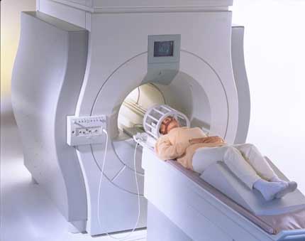
The MRI machine can be a large cylinder where a body's put in a supine position. Ahead of the procedure, metal jewelry on your body, braces, and other metal objects are removed. The sufferer is secured with straps up for grabs to minimize mobility which are more accurate results.
Special products are coupled to the head that generate and receive radio waves. There's significant noise within the device, and so the patient emerged earplugs for maximum comfort.
Analysis of the result
Inside the resulting image, you will see blood vessels, neoplasms, dense and soft tissues. The image is consumed several projections in the desired depth, thanks to that this doctor will be able to appraise the health of the area of the brain. Through the scanning process, a number of images are obtained, each of which can have a layer-by-layer portion of the cerebral tissue. With thanks to the different contrast of the same image, everything may be appreciated.
The images show: white matter, cerebral aqueduct, cerebellum, trunk, vascular structures. The tomograph creates images which might be presented as highlighted and eye shadows.
Decryption
When decrypting, a unique interpretation protocol is utilized. The pictures obtained are weighed against reference MRI data from a healthy brain. To accurately decipher the info received, the specialist has to thoroughly know the physiological and pathological anatomy. It can be obligatory to understand the peculiarities from the operation of the tomograph, which has been used for the examination.
For more information about glavnoe.ua see our new web page.