Foot Bone Spur Surgeon: Minimally Invasive Options for Bony Overgrowths
That hard, pea-sized bump you feel rubbing inside your shoe is not imaginary. It is bone, and it grew there for a reason. As a foot and ankle surgery specialist, I see this story every clinic day: a runner who cannot push off because a dorsal spur bites at the top of the big toe, a teacher whose heel lights up by lunch because a posterior calcaneal bump scrapes in the back of the shoe, a soccer midfielder who lost ankle dorsiflexion after a sprain and now jams on every cut. Bone spurs are the body’s attempt to stiffen foot and ankle surgeon NJ or protect an area under stress. When they become the problem rather than the solution, a minimally invasive foot and ankle surgeon can turn a frustrating cycle into a straightforward fix.
Why bone spurs form, and why location mattersBone overgrowth, or osteophyte formation, is the skeleton’s response to repetitive traction, compression, or joint instability. The foot is a prime target because it absorbs thousands of load cycles every day. Mechanics differ by site:
The site determines both symptoms and strategy. A top rated foot and ankle surgeon weighs not only the spur size on imaging but also the biomechanics that created it. Removing bone without addressing the underlying driver invites recurrence.
Signs that point to a bone spur rather than soft tissue painBone spur pain is usually focal, reproducible, and made worse by direct pressure or joint motion nearing end range. I have patients touch the exact spot with one finger. A hard, fixed bump that grinds against a shoe, or a sharp pinch when the ankle reaches maximum bend, is classic. Clicking and catching at the big toe joint can occur when a dorsal spur abrades the extensor tendons.
Imaging confirms the story. Weight-bearing radiographs show bony contours under load. Ultrasound is useful for dynamic tendon irritation over a spur, and for guided injections. When I need fine detail of joint shape or plan a percutaneous route, I order a CT or weight-bearing CT. MRI adds value if I suspect cartilage injury, bone marrow edema, or tendon tears, which changes the plan for a sports foot and ankle surgeon who wants to preserve joint function.
First, exhaust the smart nonoperative optionsMany spurs settle with mechanics and inflammation control. In practice, the conservative pathway is not glamorous, but it works often enough to justify patience. Shoe changes that remove the offending pressure point can be dramatic. For dorsal midfoot bumps, I ask patients to try soft or runner-style uppers rather than stiff work shoes. For Haglund-type posterior heel pain, a heel cup that lifts the calcaneus slightly can unload the back of the shoe counter. Custom orthotics and foot surgeon directed devices help when arch mechanics or forefoot overload feed the problem.
Targeted therapy matters too. A stiff big toe wants motion, so I pair joint mobilization with a stiff-sole shoe or carbon insert to limit painful dorsiflexion during daily steps while we calm inflammation. An ankle impingement pattern responds to restoring talar glide with manual therapy and balance work for instability. Anti-inflammatory measures include topical agents, short NSAID courses when appropriate, and ultrasound-guided injections. I am careful with steroid around the Achilles insertion and plantar fascia due to rupture risk. For selective cases of plantar heel pain with a spur, shockwave therapy can help, although the spur itself is not the target.
When symptoms persist beyond six to twelve weeks of good nonoperative care, or when a patient’s sport or work function is boxed in by mechanical impingement, I discuss surgical options.
How a minimally invasive foot and ankle surgeon approaches bone spursMinimally invasive does not mean minimal results. It means targeted work through small portals, assisted by imaging and specialized instruments, to remove the bony block while respecting soft tissue. The technique depends on the location:
Big toe joint dorsal spur, cheilectomy: For early to mid-stage hallux rigidus, dorsal osteophytes can be shaved through a small incision, often 1 to 2 centimeters, with a low-profile burr. Under fluoroscopy, I contour the metatarsal head and proximal phalanx so the toe clears during push-off. Done well, this restores 20 to 30 degrees of dorsiflexion in many patients and reduces shoe irritation. Literature reports pain relief and return to activity in a majority of cases, often 70 to 90 percent for grade I to II disease. Surgeons choose between open mini-incision and percutaneous techniques based on spur size and cartilage status. A big toe joint surgery surgeon will also evaluate whether a cheilectomy is enough, or whether a motion-preserving implant or fusion suits higher grade arthritis.
Dorsal midfoot exostectomy: The classic lacing bump can be removed with a percutaneous burr under fluoroscopy through a 3 to 5 millimeter portal. The midfoot skin is thin, and superficial nerves cross the field, so tactile control matters. When hypermobility or underlying arthritis drives the spur, I counsel that removing the bump alone treats the rubbing, not the joint mechanics. Rarely, a midfoot fusion surgeon may propose fusion if pain is arthritic rather than purely impingement based.
Anterior ankle impingement arthroscopy: For athletes with dorsiflexion block and spurs at the tibial lip or talar neck, an ankle arthroscopy surgeon uses two or three portals at the front of the ankle. A shaver and burr contour the spurs while preserving cartilage. The same setup allows treatment of synovitis or small loose bodies. Return to running and cutting can be quick compared to open surgery when ligaments and cartilage are healthy, with high satisfaction rates reported across multiple series.
Posterior calcaneal spur and Haglund deformity: The decision fork is critical here. If the pain is primarily from the bony bump rubbing on shoes, an endoscopic calcaneoplasty can contour the posterosuperior calcaneus through two small portals, keeping the Achilles attached. If degenerative Achilles insertion is the main driver, or there are intratendinous calcifications, a larger open approach with partial detachment and reattachment may serve better. A minimally invasive foot and ankle surgeon will explain these trade-offs. The temptation to do less must not under-treat the tendon.
Plantar heel spur: I rarely chase a plantar spur surgically unless I am addressing recalcitrant plantar fasciitis. Even then, the target is fascia release or debridement rather than the spur itself. Removing a plantar spur that is not the pain source is a common pitfall seen in foot and ankle surgeon second opinions.
Peroneal, posterior tibial, and navicular traction spurs: If a spur is secondary to tendon disease, the plan addresses the tendon first. A peroneal tendon repair surgeon or posterior tibial tendon surgery specialist may debride a small enthesophyte as part of the tendon procedure, but the real win comes from restoring tendon glide and strength.
What to expect from a surgical evaluationA board certified foot and ankle surgeon will map symptoms to structure, then test mechanics. Expect weight-bearing X-rays at minimum, possibly ultrasound at the bedside. If prior surgery failed or the anatomy is complex, a revision foot surgery specialist may order CT to model the exostosis in 3D. I measure available joint motion, palpate Have a peek here for nerve irritation, and watch gait. For runners and athletes, I ask about training volume, surfaces, and footwear history. For seniors and patients with diabetes or neuropathy, a diabetic foot and ankle surgeon also screens vascular status and skin integrity, which affects incision placement and healing timelines.
I like to show patients the spur and trace a likely resection on the image. Visualizing the amount of bone that needs to go, and the nearby nerves and tendons, helps set expectations for both the minimal incision approach and the protective period afterward. If the pain pattern hints at more than bone, such as cartilage damage or ankle instability, the plan expands. That is why a foot and ankle surgeon using arthroscopy or fluoroscopy is valuable. The tools match the task rather than forcing a one-size approach.
When to consider seeing a surgeon A hard, localized bump that rubs in shoes despite footwear changes and padding. Joint motion blocked at an end range with sharp pain, especially at the big toe or front of the ankle. Symptoms that persist beyond six to twelve weeks of focused therapy and orthotic support. Loss of sport or work function, such as inability to squat, climb, or push off the hallux. Recurrent swelling or nerve irritation over a bony prominence, or failure of prior spur surgery. Inside the operating room, outside the large incisionThe technical choreography of minimally invasive spur removal is consistent: small portals, continuous imaging, and careful protection of soft tissue. A minimally invasive foot and ankle surgeon uses burrs designed for percutaneous work, with fluid irrigation to avoid heat injury. Fluoroscopy or live ultrasound confirms position. In ankle arthroscopy, fluid distends the joint and a camera guides a burr to clean the tibial or talar lip. In hallux cheilectomy, the burr contours the dorsal metatarsal head while the surgeon cycles the toe to test clearance. When nerves cross the field, such as the superficial peroneal branch near the anterior ankle or the saphenous dorsomedially at the big toe, tactile awareness and anatomic landmarks matter more than speed.
Anesthesia is usually regional with sedation or light general. Many cases suit same day outpatient surgery. For patients with cardiac concerns, sleep apnea, or diabetes, a foot and ankle podiatric surgeon or foot and ankle orthopedic surgeon will coordinate with anesthesia to choose the safest pathway.
Recovery timelines by procedurePatients ask about recovery before anything else, which is fair. I give approximate ranges that reflect real practice, and I anchor them to milestones rather than dates alone:
Cheilectomy for hallux rigidus: Most patients bear weight right away in a stiff shoe or postoperative sandal. Swelling peaks within two weeks, then recedes over four to six weeks. Gentle motion starts within days to prevent stiffness. Many desk workers return in a week, while laborers and athletes need four to eight weeks depending on demands. Running often resumes between six and ten weeks if pain allows.
Dorsal midfoot exostectomy: Weight bearing in a boot or supportive shoe is typical. The skin is thin, so I watch for nerve sensitivity. Driving returns in a week or two on the left side, longer on the right, based on comfort and safety. Residual numbness over a small patch is possible but usually fades.
Anterior ankle impingement arthroscopy: Crutches for comfort in the first days, then rapid progression as swelling allows. Range of motion work begins early. Many athletes jog by four to six weeks and return to sport between six and twelve weeks, provided there is no cartilage injury or ligament work.
Endoscopic calcaneoplasty: Protected weight bearing in a boot for a couple of weeks is common, then transition to shoes as incision tenderness allows. If the Achilles was not detached, strengthening starts early. If tendon disease required more extensive work, the timeline extends.
Every recovery is adjusted for age, vascular health, nicotine exposure, and coexisting tendon or cartilage problems. A foot and ankle surgeon for seniors builds in more time for swelling control. A foot and ankle surgeon for athletes calibrates load progression to prevent relapse.
Risks, trade-offs, and how we prevent problemsAny surgery invites risk, even with small incisions. The complications we talk through are specific and manageable with planning.
Nerve irritation: Superficial nerves cross many target zones, which is why I map them and warn about possible temporary numbness or tingling. Permanent painful neuroma is rare. Respecting soft tissue planes and avoiding aggressive retraction matter more than the size of the incision.
Under-resection and recurrence: Removing too little bone can leave impingement, while over-resection of a stabilizing lip can destabilize a joint. Real-time fluoroscopy and intraoperative motion checks help balance this. I show patients how much bone I plan to remove and why.
Wound problems: Small portals lower the risk, but not to zero. Smoking, high BMI, diabetes, and steroid exposure slow healing. A diabetic limb salvage surgeon will optimize glucose control and offload pressure points to protect incisions.
DVT and CRPS: Blood clots are uncommon in these cases but not impossible. I screen risk factors and use mobilization early. Complex regional pain syndrome is rare but serious. Early diagnosis, vitamin C protocols in select patients, and careful pain control help reduce incidence.
Joint stiffness: Especially after big toe work. Early, guided motion avoids scar blocks. Over-aggressive early activity can inflame tissues and slow progress, which is why a measured plan with a foot and ankle surgery physical therapy team pays off.
If you are comparing options, ask a foot and ankle surgeon for chronic pain or a revision ankle surgery surgeon to explain what they do to minimize each of these risks, and how they handle them if they occur. A candid plan is part of being a best foot and ankle surgeon for your case, not a guarantee that nothing bad ever happens.
How outcomes are judged, and what “success” meansWith bone spur surgery, success is not just a pretty X-ray. It is a meaningful change in function and comfort. Patients usually want three things: less pain, better motion, and fewer shoe problems. For cheilectomy in early hallux rigidus, multiple studies report most patients achieve significant symptom relief and improved dorsiflexion, with many returning to preferred footwear and activities. For anterior ankle impingement, arthroscopy yields high rates of return to sport within a few months when cartilage is preserved. Endoscopic calcaneoplasty decreases shoe conflict and reduces posterior heel pain when Achilles integrity is good.
Where outcomes lag is when the spur is a symptom of advanced arthritis or tendon degeneration. In those cases, a foot arthritis surgeon or ankle arthritis surgeon might pivot to joint preservation or reconstructive strategies rather than simple exostectomy. That could mean osteotomy, fusion, or even joint replacement in other parts of the foot or ankle when warranted.
Choosing the right surgeon for your spurTitles vary. You will meet both a foot and ankle orthopedic surgeon and a foot and ankle podiatric surgeon with excellent outcomes. Focus on case volume for your exact problem, board certification, and whether the surgeon offers both open and minimally invasive approaches. A double board certified foot and ankle surgeon is not automatically better, but certification signals training standards. Look for a foot and ankle surgeon using advanced imaging for planning, and someone comfortable with arthroscopy and percutaneous burr techniques. If you are an athlete, a foot and ankle surgeon for runners or a sports foot and ankle surgeon who understands season timing and return-to-play metrics adds value. If you have diabetes or vascular disease, a diabetic foot and ankle surgeon who coordinates with medical specialists is critical.
Second opinions are healthy, particularly for failed prior surgery or complex deformities. A foot and ankle second opinion surgeon should explain why a procedure failed and what will be different next time. Be wary of anyone who promises cure without discussing risks or alternatives.
Practical details that affect cost, logistics, and scarringCosts vary with region, facility fees, anesthesia, and whether imaging or implants are needed. Percutaneous and arthroscopic spur work is often outpatient same day surgery, which can lower facility charges. If you want a clearer number before committing, ask for a written estimate that separates surgeon, anesthesia, and facility fees. Many practices can give a range based on CPT codes once the plan is set. Insurance authorization matters, particularly if advanced imaging such as CT is required preoperatively.
Scarring after minimally invasive spur removal is usually minimal. Still, portal care and scar management count. I recommend gentle massage once healed, silicone gel sheeting when incisions are closed, and protection from sun for at least six months to reduce darkening. For patients prone to keloids, we discuss strategies early.
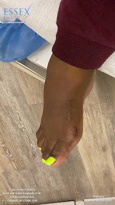
A day in the life of minimally invasive spur surgery Arrival and marking: The surgeon marks the tender area and draws safe corridors that avoid nearby nerves. Imaging is reviewed one last time. Anesthesia and positioning: You receive regional anesthesia with sedation or general, depending on the case. A tourniquet may be used briefly to limit bleeding. Portal creation and resection: The surgeon creates a tiny incision, protects soft tissue, then uses a burr or arthroscopic shaver under live imaging to contour the spur. Range of motion is tested to confirm clearance. Closure and dressing: One or two sutures close each portal. A compressive dressing limits swelling. You receive a protective shoe or boot. Early recovery: You are encouraged to elevate and move within the plan. Most patients go home the same day, with a simple pain regimen and follow-up in 10 to 14 days. Edge cases, and when less is not moreSometimes the smallest incision is not the smartest choice. If a dorsal midfoot spur hides an unstable Lisfranc joint, removing the bump without stabilizing the joint sets you up for persistent pain. If a posterior heel spur lives within a degenerated Achilles, endoscopic bone work without tendon treatment often fails. A complex foot reconstruction surgeon knows when to combine procedures or recommend a staged plan.
Similarly, a patient with significant ankle arthritis and large anterior osteophytes may do poorly with pure arthroscopic debridement. An ankle deformity correction surgeon or ankle fusion surgeon might offer osteotomy or fusion when joint preservation is no longer realistic. For focal cartilage defects in the ankle, an ankle cartilage repair surgeon or osteochondral lesion ankle surgeon may combine spur work with microfracture, drilling, or grafting to protect long-term function.
Rehabilitation that respects biologyThe best surgical plan fails without good rehab. Early on, the goals are swelling control, protected motion, and gait normalization. I work with therapists who see foot and ankle problems daily. For hallux procedures, we emphasize gentle dorsiflexion without forcing pain, scar glide, and intrinsic foot strength. For ankle impingement, we progress dorsiflexion mobility, peroneal activation, and proprioception. For posterior heel cases, we protect the Achilles first, then build eccentric strength on a schedule that matches tendon healing, not calendar impatience.
Patients who do well take ownership of the simple things: consistent elevation in the first week, shoe choices that respect the incision, and adherence to the progression rather than jumping ahead on a good day. A foot and ankle surgeon for chronic ankle sprains or tendon tears will often coordinate a return-to-run or return-to-work protocol with clear checkpoints to prevent surprises.
Final thoughts from the clinicBone spurs in the foot and ankle are common, visible, and fixable when they are the true pain source. The trick is to match what you feel under your fingertip to the mechanics that created it, then choose the least invasive method that actually solves the problem. In my practice, that often means percutaneous cheilectomy for a dorsal hallux bump, arthroscopic debridement for anterior ankle impingement, or endoscopic calcaneoplasty for a pure Haglund conflict. When the story is more complicated, the solution expands appropriately.
If you are evaluating your options, start with a focused assessment from a board certified foot and ankle surgeon who treats your exact issue weekly. Ask to see the spur on your imaging, hear the plan in plain terms, and review the alternatives. With the right match between problem and technique, minimally invasive spur surgery can trade a stubborn daily rub for a small, quiet scar and a return to the way you move when bone is not in the way.