Enzymes Present In The Sperm

🔞 ALL INFORMATION CLICK HERE 👈🏻👈🏻👈🏻
Enzymes Present In The Sperm
Male mice were injected i.p. with 2.5 mg/kg mitomycin C, 100 mg/kg ethyl nitrosourea or saline and mated with untreated virgin females five weeks later. Sperm from 64 of the F1 male progeny were analyzed histochemically for acrosin, succinic dehydrogenase and alpha-glycerophosphate dehydrogenase activity. The frequency of F1 males with sub-normal sperm enzyme activity was significantly higher among progeny from treated males than in controls. These results show that analysis of sperm enzyme activity in F1 males is a practical method for detection of transmitted mutations induced in a treated parent.
Content uploaded by Brahma B. Panda
Content may be subject to copyright.
Experientia 44 (1988), Birkh/iuser Verlag, CH-4010 Basel/Switzerland 253
Sperm enzyme activity analysis in individual sperm for detection of heritable mutations in mammals
B. B. Panda ~, G. Ficsor 2 and L. C. Ginsberg
Department of Biology and Biomedical Sciences, Western Michigan University, Kalamazoo (Michigan 49008, USA)
Received 19 May 1987; accepted 26 October 1987
Summary. Male mice were injected i.p. with 2.5 mg/kg mitomycin C, 100 mg/kg ethyl nitrosourea or saline and mated with
untreated virgin females five weeks later. Sperm from 64 of the F~ male progeny were analyzed histochemically for acrosin,
succinic dehydrogenase and alpha-glycerophosphate dehydrogenase activity. The frequency of F~ males with sub-normal
sperm enzyme activity was significantly higher among progeny from treated males than in controls. These results show that
analysis of sperm enzyme activity in F ~ males is a practical method for detection of transmitted mutations induced in a treated
Key words. Mice; heritable mutations; sperm enzyme activity.
Mammalian germ cell tests are important for human genetic
risk assessment 3+ 4. We report a germ cell test in which en-
zyme activity is evaluated in individual sperm of F 1 male
mice from treated or control sires. We found that the muta-
genicity of ethyl nitrosourea (ENU) or mitomycin C (MC) is
detected by this method. HA (ICR) F o males were injected
once i.p. with 2,5 mg/kg MC, 100 mg/kg ENU or saline vehi-
cle. Five weeks later each male was caged with two virgin HA
(ICR) females for one week. After mating, the males were
discarded and the females individually caged. The F~ males
were either killed by cervical dislocation or were semicastrat-
ed under ether anesthesia to obtain sperm samples.
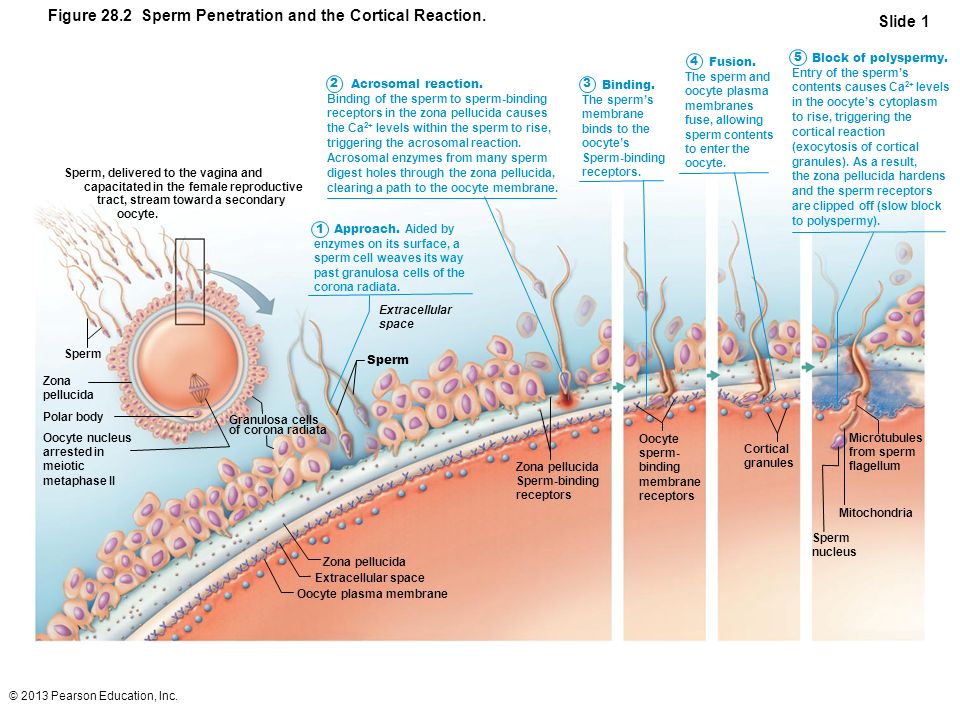
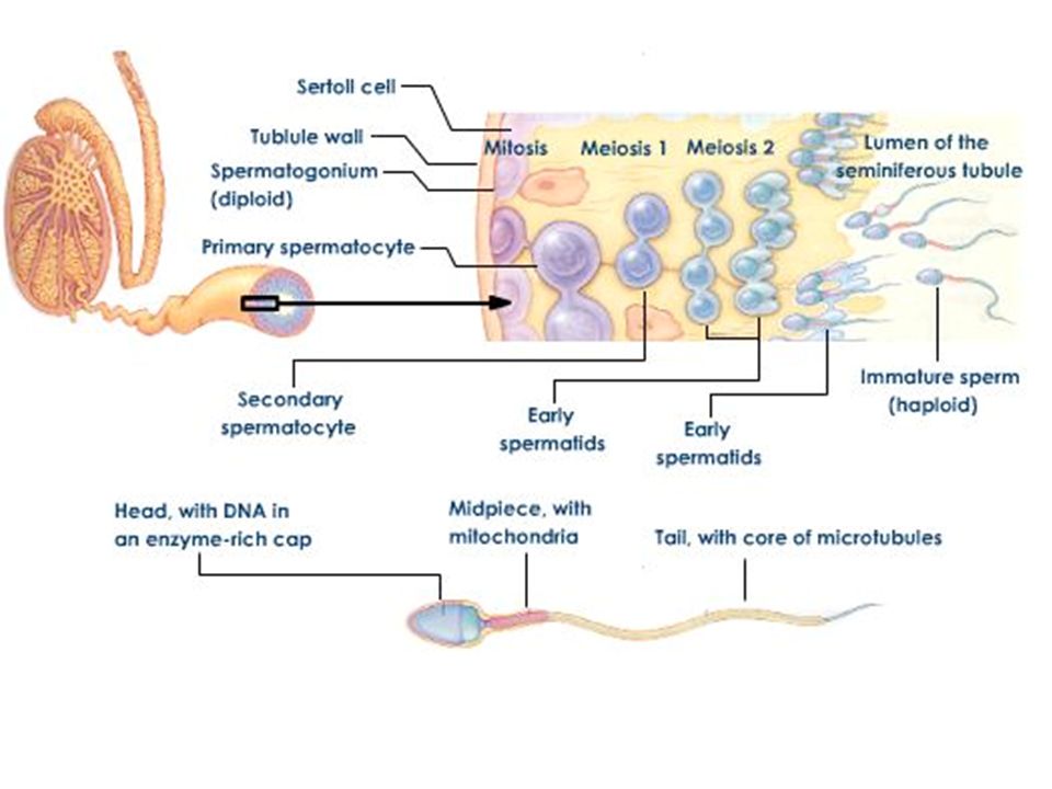
The sperm from each F~ male was used for histochemical
detection of acrosin, succinic dehydrogenase (SDH) and
alpha glycerophosphate dehydrogenase (AGPDH) activity.
At least two microscope slides were made from each F~ male
and for each assay. The slides were randomized and blindly
scored. 250 sperms were examined at x 400 magnification
from each Fa mouse and for each assay. A sperm was scored
as positive for acrosin activity if its head was surrounded by
a yellow halo in a dark blue background 5, 6. Sperm without
acrosin activity lacked a halo. Both SDH and AGPD are
located in the mid-piece region of the sperm. When active,
both enzymes produced the reduced violet precipitate of
paraiodonitrotetrazolium violet localized in the mid-piece
region 6, 7. Sperm without SDH or AGPD activity lacked the
Two successive statistical analyses were performed: First, the
number of enzyme inactive sperm in each F~ male was com-
pared to similar numbers in F~ males from control sires. If
enzyme inactivity in F1 males exceeded the 99.9% confi-
dence interval of the control mean such male was classified
as a variant male in (fig., open shapes). Second, the statistical
significance of differences between the number of variant F1
males from treated or control sires was determined using
homogeneity chi-square test on a 2 x 2 contingency table 11.
The figure shows the number of F~ mice with percentage of
inactive sperm for acrosin, SDH and AGPDH in individual
F~ males from treated or control sires. Generally more vari-
ant males were present among F 1 males from treated sires
than in controls. The statistically significant effect of treat-
Judging from the high frequency of Fa variant males from
treated sires, we conclude that mutation of many different
genes may result in loss of enzyme activity. A large genetic
target, as might be the case with this test, means that fewer
F1 animals are required (only 64 in this study) to detect a
Sperm enzyme inactive variant F1 males from treated or control sires a
Agents tested Dosages Variant F1 Males/total F~ Males b
in F o males (mg/kg) Acrosin SDH AGPDH
a 24 F 1 males from ENU-treated sires reached sexual maturity. Only 23
of these had enough sperm for statistical evaluation for acrosin activity
and 20 each for SDH and AGPD activity, b Significantly different from
controls at* p _< 0.05 and** p < 0.01.
Percentage of sperm without acrosin, succinic dehydrogenase or alpha-
glycerophosphate dehydrogenase activity in F 1 sons sired by control,
ethyl nitrosourea or mitomycin C treated F 0 male mice. The enlarged
FROM FoCONTROL FROM F o MC FROM F o ENU
open shapes represent mice with inactive sperm that exceed 99.9 % confi-
dence interval of the control mean.
This chapter reviews historical and current trends in the evaluation of a male reproductive toxicant. This evaluation encompasses the design, conduct and interpretation of studies evaluating alterations in the structure and function of male reproductive organs. While the primary objective of any reproductive toxicity study is to quantitate reproductive outcome, newer investigators are often faced with challenging questions regarding the choice of endpoints and thus some of the important aspects of study design are discussed. The first half of this chapter discusses standard approaches and regulatory guidelines governing the timing and design of these studies. The latter half of the chapter discusses current methodologies for evaluating effects on male reproductive health, including functional assessments of spermatogenesis and fertility, along with gross macroscopic and microscopic evaluations. In addition, toxicant-induced effects on the developing male and aspects of male-mediated developmental toxicity are presented. Finally, data interpretation and the importance of evaluating the weight of the evidence are emphasized in the evaluation of a male reproductive toxicant.
Kothare and DeSouza (5) have reported the cytochemical distribution of succinic dehydrogenase in normal and abnormal spermatozoa using p-nitrophenyl substituted ditetrazole. They found the enzyme located in the middle piece and the head, but absent in the neck and tail. Thin sectioning studies have shown a complete absence of mitochondria in the head of the human spermatozoa (1). In view of this ... [Show full abstract] observation, and the close spatial association between this enzyme and mitochondria (2, 3, 7, 9) it seemed desirable to examine the exact localization of succinic dehydrogenase in the human sperm at the magnifications available in the electron microscope.
July 1971 · Canadian Journal of Physiology and Pharmacology
An injection of a single dose of cortisone acetate (5 mg/100 g body weight) to 9-day-old rats resulted in the following changes in brown adipose tissue 24 h later: (1) the fresh weight was increased due to fat accumulation; (2) the DNA content of whole interscapular brown fat stayed unaltered, while the RNA content was increased; (3) specific activities of cytoplasmic alpha-glycerophosphate ... [Show full abstract] dehydrogenase and malic enzyme were increased; (4) the percentage of mitochondrial protein in the whole tissue protein was not changed, but mitochondria seemed to be more fragile, fewer were recovered by a standard isolation procedure, and more cytochrome c oxidase contaminated the microsomal fraction; (5) mitochondrial alpha-glycero-phosphate dehydrogenase and succinate dehydrogenase activities were decreased per milligram homogenate protein and (in isolated mitochondrial fraction) per milligram mitochondrial protein; (6) the endogenous respiration of brown fat mitochondria was activated much less by carnitine and CoA; and (7) CO2 formation from palmitate-14C by isolated mitochondria was considerably lower.A similar injection to 30-day-old rats had no significant effect.It is suggested that a single injection of cortisone affects the mitochondrial structure of brown adipose tissue and the ability to oxidize fatty acids and that it is effective on day 10 but not on day 30.
February 1970 · Histochemie. Histochemistry. Histochimie
February 1972 · Zhurnal nevropatologii i psikhiatrii imeni S.S. Korsakova (Moscow, Russia: 1952)
© 2008-2021 ResearchGate GmbH. All rights reserved.
Enzyme - Wikipedia
(PDF) Sperm enzyme activity analysis in individual sperm for detection...
The lytic enzyme present in semen is
Enzymes Present In The Sperm | PsyLan.ru | ВКонтакте
what is the enzyme present in acrosome of sperm - Brainly.in
The lytic enzyme present in semen is
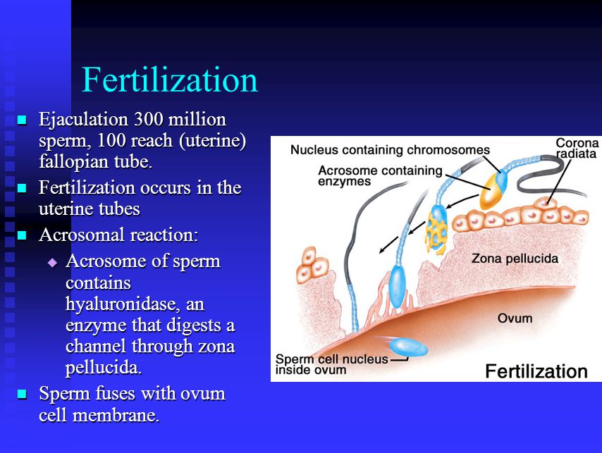
The fructose provides energy for the sperm after they are ejaculated. Prostaglandins stimulate contractions of the uterus, which help move sperm up the female reproductive tract. The single prostate gland secretes an alkaline fluid containing calcium, citric acid and enzymes like the lytic enzyme hyaluronidase. The prostatic fluid may be important in neutralizing the acidic environment of the vagina and in increasing sperm cell motility. During sexual arousal, the paired bulbourethral glands, located on each side of the urethra, release a mucous secretion.
Semen consists of which of the following enzymes?
Customize assignments and download PDF’s
Take Toppr Scholastic Test for Aptitude and Reasoning Win exciting scholarships and plan a great education plan Register for free
Stay upto date with our Newsletter!
Stay informed, stay ahead. Be a topper
There is a glucose sugar in the semen.
Watch learning videos, swipe through stories, and browse through concepts
Overwatch Sexy Ass
Www Porno Sex Pickup Czech
Model Double Penetration
Korean Softcore Movies
Outdoor Discount