Cardiac cycle and heart sounds 🫀
Ninja Medical Squad ❤️Cardiac cycle: It means the mechanical events occurring in the heart within one beat (from the beginning of heart beat to the next one)
HR 75 bpm ➡️0.8 sec
و العلاقة بينهم عكسيه كل ما زاد HR قل ccd cardiac cycle duration
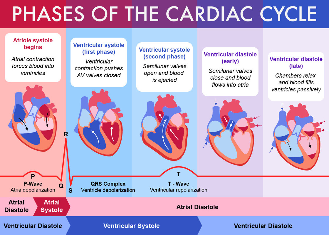
💢Cardiac cycle 8 phases
وكل phase هنتكلم فيها عن 8 نقط
1 )artial pressure
2)aoric valve
3)aortic pressure
4)AV valve
5)venticular pressure
6)venticular volume
7)HS
8)ECG
دلوقتي هنخش على phase 1
🔸️Phase(1) artial systole phase (0.1 sec)
1 )Atrial pressure (AP): ↑ few mmHg due to atrial contraction. Then returns back to zero by the end of this phase due to evacuation of atrial blood
هيزيد في الأول بعد كده هيقل لما ينزل لل venticules
2)aortic valve: closed
3)aortic pressure : decrease
علشان ال دم بيروح من الaorta لباقي الجسم
4)AV valve : open. Ventricles الدم نازل لل
5)venticular pressure: increase then decrease
في الأول بينزل من ال atrium وحجم ال Ventricles زي ما هو بعد كده حجمه بيزيد فالضعط بيقل
6)venticular volume : increases
7)HS : 4th heart sound
8)ECG : P wave
🔸️ Phase2 isometric contraction phase (0.05 )
Isometric: without any change In length of fiber muscle
1 )artial pressure : increase due to ballooning of av cups towards the atrium by sudden increase in intravascular pressure
هيزيد شوية علشان الدم في الventircles هيزيد و هيضغط على av valve فجأة فهيضغط علىatrium
2)aoric valve still closed
3)aortic pressure : still decreasing and at the end of the phase venticular pressure will be higher than aortic pressure
4)AV valve : suddenly closed
5)venticular pressure : increase
بقى أعلى من الatrium
6)venticular volume : no change
7)HS : 1st heart sound
8)ECG : qrs complex
🔸️ Phase 3 Maximum ejection phase (0.15 sec )
1 )artial pressure : decrease av valve هيرجع زي ما هو
بعد كده gradual increases علشان الدم الجاي من ivc و svc
2)aoric pressure : increases to 120 mmHg because pumped blood by ventricles
3)aortic valve :open
4)AV valve: closed
5)venticular pressure : increase from 80 to 120
6)venticular volume : decrease due to ejection of blood to aorta
7)HS : first sound continues
8)ECG : origin of t wave stars somewhere in this phase
🔸️ Phase4 reduced ejection (0.1)
1 )artial pressure gradual increases due to venous return
2)aoric valve : open
3)aortic pressure : decrease because the blood is distributed to the body
4)AV valve : still closed
5)venticular pressure : decrease becomes decreased force of pumping blood to aorta
6)venticular volume : still decreasing
7)HS : no hs
8)ECG : top of t wave
🔸️ Phase 5 protodiastolic phase (0.04) مهمة
1 )artial pressure : gradual increases
2)aortic valve : open
3)aortic pressure : aortic valve هيفضل باصص على
Sudden closure AO valve cause sharp decrease the aortic pressure called
(Incisura ) (dicrotic notch )
4)AV valve : still closed
5)venticular pressure : decrease till become lower than aorta
6)venticular volume : no change
7)HS : no hs
8)ECG : t wave continues

🔸️ Phase 6 isometric relaxation phase (0.06)
1 )artial pressure : gradual increases
2)aortic valve : suddenly closed
3)aortic pressure : decrotic notch then decrotic wave (sudden increase) due to elastic recoil of aorta
4)AV valve : still closed
5)venticular pressure : rapidly decrease
6)venticular volume : no change
7)HS : 2nd hs
8)ECG : t wave ends during this wave

🔸️ Phase 7 maximum Filling phase (0.1)
1 )artial pressure : increase then decrease due to rush to the ventricles
2)aoritc valve : closed
3)aortic pressure : decreasing
4)AV valve : opened
5)venticular pressure :around zero due to progressive venticular relaxation
6)venticular volume : increases rapidly
7)HS : 3rd is abnormal due to rapid venticular filling
8)ECG : no waves (isoelectric)
🔸️Phase8 reduced Filling phase (0.2 sec )
1 )artial pressure : around zero but higher than the ventricles
2)aortic valve : closed
3)aortic pressure : still decreasing due to moving blood to tissues
4)AV valve : open
5)venticular pressure : around 0 but lower than atria
6)venticular volume : increases
7)HS : no hs
8)ECG : beginning of p wave next cycle
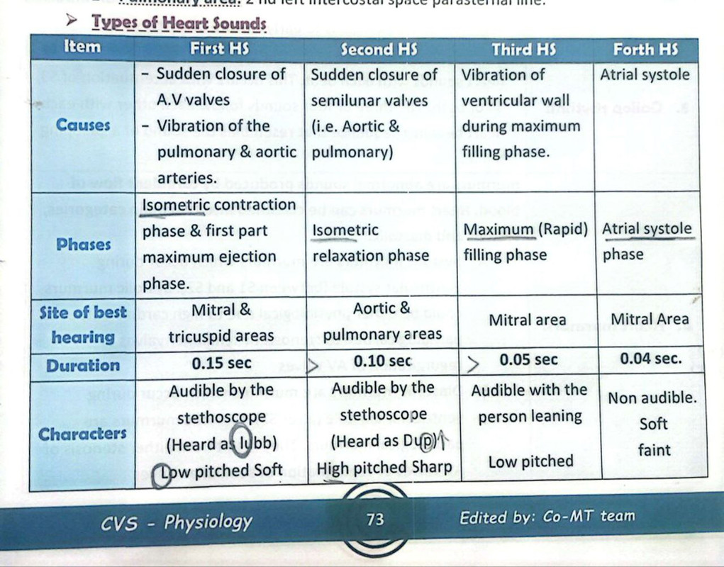
💢 The Heart sounds
4 heart sounds recorded by phonocardiography
Only 2 are heard and the third could be heard in children
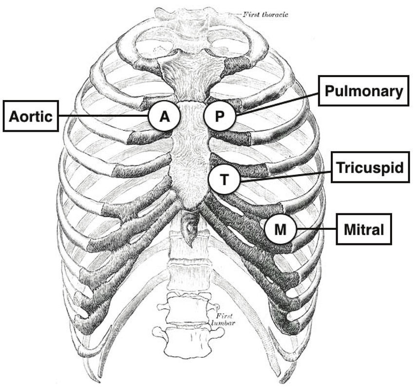
Method : stethoscope
علشان نسمع لازم نحط السماعة على asculatory areas
Mitral left 5th intercostal space mid calvicular line (apex)
Tricuspid lower part of sternum
Aortic 2nd right Coston sternal junction
Pulmonary 2nd left intercostal space
💢Types of heart sound 🩺
💢Abnormalities of heart sound
1) splitting of first Heart sound: delay in Tricuspid valve closure after Mitral valve closure (right bundle branch block )
2) splitting of second heart sound :
Physiological: delay of closure in Pulmonary valve after aortic valve
Pathological: split disappears during expiration لو استمرت
يبقى فيه Pulmonary stenosis
Which prolong ejection time of RT ventricle or early closure of aortic valve due to Mitral regurgitation الصمام مش عارف يقفل
3) gallop rhythm: more than two audible sounds heart sound
S3 or S4 شبه صوت الحصان
4)heart murmur: اضطراب في الصوت due to turbulent flow of blood
Systolic : during venticular Systole (between s1 and s2)
Physiological : high cardiac output
Pathological: stenosis of semilunar vaves
Regurgitation of av valve
Diastolic: during venticular diastole (after s2)
Pathological only due to stenosis of av valve or Regurgitation of semilunar valve .
💫💫الحمدلله الذي هدانا لهذا و ما كنا لنهتدي لولا أن هدانا الله 💫💫