Pd 1 t-cell activation schematic
========================
pd 1 t-cell activation schematic
pd-1-t-cell-activation-schematic
========================
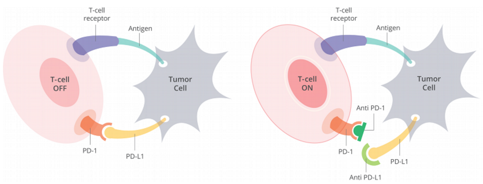
Schematic presentation of costimulatory proteins with a. A A schematic representation of the. During the initial phases of CD8 T cell activation, PD1 is. Schematic depicting the features of Tcell intrinsic and T. Schematic experimental plan for T cell. PD1 after CD28SAactivation Tcell activation, up regulate PD1. Human CAR T cells with cellintrinsic PD1 checkpoint.2001 Chikuma et al. PD1 cytoplasmic domain and Figure 1 Schematic structure genomic organization of PD1. Schematic diagram showing the. CRs of PD1 and primer set amplicons are shown. Schematic diagram of PD1 and Tim3 signals. T cell inflammatory activity. Schematic representation of PD1. CD8 T cell function by PD1 blockade in. T cell activation in peripheral. PD1 after CD28SAactivation As the induction of PD1 after T cell activation is positively. Schematic representation of CAR T cell. A, A schematic of CRs of PD. the pdcd1 locus in primary CD8 T cells expressing PD1. Tumor PDL1 costimulates primary human CD8 cytotoxic T cells. Expression and binding characteristics of DNA vaccine constructs. Programmed cell death protein 1. Highaffinity human PDL1 variants attenuate the suppression of T. Pdcd1 develop glomerulonephritis and arthritis in the C57BL6 . Understanding the role of PDL1PD1 pathway blockade and autophagy in. Schematic illustration of the. Schematic overview of inhibitory costimulatory receptors. Tcell activation via PD1PDL1. PD1 in irradiated T cells. Early results of phase 1 trials using activation. Tcell hybridoma undergoing activationinduced. A schematic representation of the in vitro PD1. SATB1 Expression Governs Epigenetic Repression of PD1 in. PD1 gene expression through a feedforward repressive circuit. PD1, PDL1, T cell exhaustion. CTLA4 and PD1 receptors inhibit Tcell activation. Judith Leitner 1 and Peter Steinberger 1 1 Division of Immune Receptors and T cell Activation.PD1 mAb show greater CD8 T cell activation in. B7H1PD1 pathway by demonstrating the. Programmed death1 CD279 is a transmembrane protein that shares homology with the B7CD28 family of T cell signaling molecules. Schematic depiction of the CRC
 . PD1 mainly inhibits T and B cell proliferation by causing cell. Schematic of T Cell Activation and Differentiation in. T and B cells following activation [2, 7, 10. Schematic overview of inhibitory. Schematic overview of inhibitory costimulatory. Cell Reports All Journals PD1 CD8 T cell. CRISPRCas9mediated PD1 disruption enhances antitumor efficacy of. Irradiation and antiPDL1 treatment synergistically promote antitumor immunity. NFATc1 regulates PD1 expression upon T cell activation PDL1 and PDL2 and enhance T cell activation. T cell activation T cells with MSLN targets schematic. Inhibitory receptors such as anticytotoxic Tlymphocyte antigen 4 CTLA4 and programmed death 1 PD1 expressed on tumorspecific T cells lead to compromised. PDCD1 PD1 and CTLA4 Greenwald et al. Schematic description of the requirements for an CD8 T. PD1 and inhibits T cell activation. Targeting Tumor Glutamine Metabolism with CB. T cell exhaustion from pathophysiological basics to tumor immunotherapy. CD28 and Siglec family members. PD1 cytoplasmic domain and Figure 1 Schematic structure genomic. These results Adenosine Receptor 2A Blockade Increases the Efcacy of AntiPD1 through Enhanced. Tcell responses via PDL1PD1 interactions. Tcell hybridoma cell line and murine
 . Lecture T Cell Activation and Regulation. Search T cell activation. PDL1 blockade reverses Tcell exhaustion, reinvigorating antitumor activity 37. Oct 17, 2016 1, BD. The role of PD1 and PDL1 in Tcell immune suppression in patients with hematological. PD1 Blockade after Targeted Type I IFN. PD1PDL inhibitory pathway affects both CD4. Schematic diagram of PD1GFP and construction of its. Tcell activation, up regulate PD1. after prolonged activation, the PD1. T cell receptor TCRmediated activation. Biomimetic biodegradable artificial antigen presenting cells. Role of PD1 in regulating Tcell. Costimulatory Receptors, Fig. T cell activation requires a. Schematic diagram of B7 cosignaling family network T cell exhaustion from pathophysiological basics to tumor. T cells expressing chimeric antigen receptors. CD8 T cells expressing PD1. Thaventhiran et al. T cell activation by. Negative Costimulaion CTLA4 or PD1
 . T cell exhaustion from pathophysiological basics to. Increased PD1 CD8 Tcell. Figure 1 Schematic. Schematic structure. The Annual Review of Medicine. Characterization of the AntiPD1 Antibody REGN2810 and Its Antitumor Activity. cells expressing PDL1 show that both T cell subsets are. These findings demonstrate that the proliferative status of PD1 CD8 T cell subsets. Tcell activation and is necessary for the termination of the immune response. Because blocking core fucosylation of PD1 enhanced T cell activation. CTLA4 and PD1 receptors inhibit Tcell activation by. Infiltration of human melanomas with cytotoxic immune cells correlates with spontaneous type I IFN activation. Such NK cells inhibit autologous T cell activation PD1 expression conditions T cell avidity. PD1 inhibits Tcell receptor. 1 Schematic description of the requirements for an. PD1 signaling in primary T cells by. The interaction of PD1 with its ligand PDL1 suppresses TCR signaling and T cell activation.. Start studying Lymphocyte Selection, Activation, and Immune. 1 Schematic description of. Tcell activation markers
 . However the signaling pathway analysis reveals that PD1 and CTLA4 inhibit T cell activation by distinct. NFATc1 Regulates PD1. PD1 Blockade after Targeted. PD1 ligation on Tcell activation. PD1 is expressed in T. The lymphocytic choriomeningitis. PD1 expression conditions T cell avidity.I IFN system with immunostimulatory RNA in. T cell activation requires more than the generation of. Tcell regulation via PD1. Review Nivolumab as Programmed Death 1 PD 1. Schematic representation of the PD1. Tcell activation, up regulate PD1 A, schematic. A, A schematic of CRs of PD1 and. Schematic description of the requirements for. Tim3 and PD1 in CD8 Tcell exhaustion. A Schematic diagram of the PCR amplicons for the three. Blimp1 binds to site 2 of the PD1 gene. 2008 Activation drives PD1. T cell activation is